WOUND HEALING
Wounds heal in one of two possible ways: regeneration or scar formation. In regeneration, the tissue that has been destroyed or damaged is replaced by tissue of the same type. This is the preferable way for wounds to heal because it preserves proper functioning of the injured site and its normal appearance. In scar formation healing, the lost tissue is replaced by fibrous scar tissue, which does not have the same properties as the original tissue and is unable to carry out the same functions (WOCN, 2022).
One of the main factors controlling how a wound heals is the depth of the wound. Shallow wounds that encompass only the epidermis and a portion of the dermis are capable of healing by regeneration because the damaged tissue can reproduce itself. However, with deeper wounds in which multiple tissue layers are involved, including subcutaneous tissue down to muscle, tendons, and bone structure, regeneration is not possible. These structures are unable to reproduce themselves, and the only option for healing is scar tissue formation.
Phases in Wound Healing
The body has a sequential mechanism to heal acute wounds through regeneration. A noncomplicated surgical incision, for example, will go through the following healing process:
- Hemostasis
- A short inflammatory phase
- Proliferation
- Maturation
HEMOSTASIS
Hemostasis begins immediately after a wound occurs, and it is initiated by blood coming in contact with the collagen in the tissues. This triggers what is called the clotting cascade. The purpose of hemostasis is to stop bleeding. Activated platelets in the wound bed release platelet-derived growth factors, which control and hasten the healing process. Activated platelets are also involved in creating the fibrin structure that leads to the formation of a fibrin clot, which stops the bleeding. The fibrin clot also serves as an initial matrix within the wound area into which cells can transfer. This process of clot formation happens rapidly if there are no bleeding abnormalities. Individuals with an impaired clotting mechanism will experience an impaired healing process (Baranoski & Ayello, 2020; Shah et al., 2018).
When larger blood vessels are severed in an acute wound, further measures to stop bleeding are usually needed, such as the application of manual pressure, cauterization, or suturing.
INFLAMMATION
Once a clot is formed and bleeding ceases, the inflammatory phase begins. The inflammatory phase is a normal and essential part of wound healing that establishes a clean wound bed. The platelet-derived growth factors attract white blood cells to the wound. The first white blood cells on the scene—polymorphonuclear cells, also called neutrophils—are the initial line of defense; their function is to remove bacteria from the wound through enzymatic activity.
The various biologically active molecules being released into the wound also hypersensitize the endings of local pain nerves, causing them to react to smaller amounts of chemical and mechanical irritation, thus making the wound site tender. Together, these processes produce local inflammation.
The number and activity of the neutrophils decline as the inflammatory process continues, and by the third day of wound healing, macrophages are the predominant white blood cells in the wound. Macrophages are scavengers that continue to debride (or cleanse) the wound biologically by removing dead and dying bits of tissue, dirt, and bacteria. Macrophages, which are derived from tissue monocytes, are an essential component of the initial phases of wound healing.
A decreased level of macrophage activity in the wound is associated with prolonged and delayed wound healing. Individuals with uncontrolled diabetes and diabetic wounds are noted to have low macrophage counts and difficulty with wound healing. Macrophages also release growth factors, chemicals that stimulate the growth of fibroblasts, endothelial cells, and epithelial cells, all of which quickly transition the wound into the proliferative phase of healing.
It is important for the clinician to recognize that induration, warmth, redness, and swelling are normal findings during the inflammatory phase of wound healing and are not, at this stage of the process, an indication of wound infection. It is also good practice to share this information with the patient (Baranoski & Ayello, 2020).
PROLIFERATIVE PHASE
The next set of events in wound healing constitute the regenerative, or proliferative, phase. This phase begins when fibroblasts (the cells responsible for the synthesis of the new connective tissue) are attracted to the wound by growth factors and white blood cells. Fibroblasts are the only cells capable of synthesizing connective tissue, and it is important to note that they can be damaged by certain antiseptics.
In acute wounds, collagen fiber production normally begins around the fifth day after injury. Collagen is a structural tissue protein found in various forms throughout the body. Collagen fibers are composed of the protein collagen, which is the most frequently occurring protein in the human body. Collagen provides strength and support to connective tissue, and adequate collagen production is an essential part of tissue repair and wound healing. At the same time, new blood vessels are growing into the wound.
Presently, 29 separate collagens have been isolated in vertebrate tissue. At a minimum, eight distinct collagens are present in human skin. Type 1 collagen is the most common type of collagen and is the principal collagen found in the human dermis. Along with type 111 collagen, it forms wide extracellular fibers in the dermis (Calonje, 2020).
Together, the newly forming cells, blood vessels, and loose extracellular matrix are called granulation tissue. Granulation tissue fills the base of an open wound. Healthy granulation tissue contains newly growing blood vessels and should be beefy red with a bumpy, uneven surface resembling velvet.
MATURATION
Maturation, sometimes called remodeling, is the last stage of wound healing. This phase can last up to one year after the wound occurrence and is characterized by strengthening, defining, and debulking of the final scar tissue.
A wound that heals without complications will achieve 80% of its normal tensile strength. Tensile strength refers to the skin’s ability to resist breakdown under tension, and it is a very important factor in maintaining normal skin integrity. It will never regain 100% tensile strength, something for clinicians to keep in mind with caring for patients with a healed wound, especially with healed pressure ulcers/injuries. This lack of regular tensile strength makes these areas more prone to further wound development.
The above phases of wound healing are usually discussed as separate entities, but in reality, wound healing is an intricate process with overlapping phases.
THE HEALING PROCESS AND CHRONIC WOUNDS
A chronic wound will not move through the healing process described above. Research into why this occurs includes the study of the molecular and cellular changes that happen in the conversion of an acute wound into a chronic one. These research findings are the basis for new and innovative therapies that seek to selectively correct the abnormalities that impede wound healing.
Whereas acute wounds are found to have high levels of growth factors, these are markedly decreased in chronic wounds. One important discovery is that acute wounds that are subjected to frequent episodes of injury can evolve into chronic wounds.
What has been discovered is that chronic wounds stall in the inflammatory phase of wound healing because of the presence of free-flowing (planktonic) bacteria in the wound and the formation of biofilm. Biofilm is defined as groups of microorganisms (bacteria and fungi) that attach themselves to a surface and become entrenched in a hydrated matrix composed of an extracellular polymeric substance (Baranoski & Ayello, 2020).
The symbiotic relationship between the bacteria in the biofilm allows their collective strength to create a formidable barrier to attack, and their densely packed matrix does not readily allow penetration by white cells and antibodies.
Research has also found that the bacteria found in the center of the biofilm become dormant and produce no metabolic activity; this greatly increases their resistance to antibiotic therapy, since antibiotics attack actively dividing bacteria. This provides the biofilm with a high level of immunity to standard treatment that readily kills planktonic bacteria, and it presents one of the major challenges in chronic wound care—the eradication of biofilm and preventing it from regrouping. (See also “Biofilm and Infection” later in this course.)
Scar Tissue Formation
Scar tissue formation is one of the two ways in which wounds heal, and all full-thickness wounds will form scars. There are noticeable differences between scar tissue and normal skin. There is less elasticity in scar tissue; it has fewer blood vessels, resulting in a decreased blood supply; and it appears lighter in tone than the surrounding skin.
Scars are the natural patches produced in a healing wound. They are the end product of the wound healing process and have diminished strength compared to normal tissue. Even years after a wound heals, it has been found that scar tissue never fully recovers the strength of normal tissue (Baranoski & Ayello, 2020).
In the first few days after an injury, closed skin wounds are being knit weakly together by the forming scar tissue. By about day five, the basic architecture of the wound patch has been established, and from then on, the healing process consists largely of strengthening and remodeling the scar.
Scars can take six to nine months to mature. New scars tend to be red and thick for a month or two before gradually becoming less vascular (i.e., paler), less bulky, and flat. It can take as long as five years for a scar to reach its final color.
MINIMIZING SCARS
The width of the scar can be minimized by:
- Thorough debridement
- Careful suturing (avoiding inversion of the skin edges)
- Removing excess granulation tissue
- Good secondary wound care (especially keeping the wound from becoming infected)
- Removing sutures promptly
PROBLEM SCARS
Scars are a natural result of healthy healing, although scars are imperfect replacements for damaged tissue. Normal scars can lead to problems. Even under the best healing conditions, some normal scars may end up interfering with the movement of the skin and the underlying tissue. In addition, some normal scars are unsightly.
When the healing situation is not ideal, scars are more likely to become problems. After poor healing, some scars become unnecessarily large or unnecessarily weak. For example:
- Infections, tissue necrosis, sebaceous skin, and wounds perpendicular to natural lines of minimal skin tension will all lead to scars that are larger than normal.
- If a wound reopens before it is effectively sealed (called dehiscence), the scar will be wider and usually weaker.
- If too few capillaries grow into the forming scar tissue, leading to ischemia, the scar will be very weak and may develop into an ulcer.
The wound patching process may also go overboard and generate too many new cells or, more commonly, too much collagen in the scar. Such scars will enlarge and bulge from the wound. Scars built of too many cells (mainly fibroblasts) are called desmoids or aggressive fibromatoses. Scars built from too much collagen are either hypertrophic scars or keloids. When excessive scars form tight ridges along the skin and permanently interfere with normal movement, they are called contractures.
Keloids
Keloids are benign tumors that grow beyond the bounds of a wound and do not regress. Keloids are caused by the excess deposition of collagen in a healing wound. The tendency to form keloids is genetic, and there are, at present, no preventive measures. Patients with darkly pigmented skin are particularly susceptible.
Unlike hypertrophic scars (discussed below), keloids develop late in the healing process; they can show up months or even years after the injury. Keloids bulge out beyond the edges of the wound, and some keloids can become sizeable. The typical presentation of a keloid is as a raised, hyperpigmented nodule that is firm to the touch. Keloids that do not regress spontaneously are usually found on the upper half of the body.
Successful treatment of keloids is challenging. Treatments that have shown some success include corticosteroid injections, chemotherapy, radiation, lasers, and surgical excision. However surgical removal of the keloid is not normally performed unless the tissue becomes pendulous (WOCN, 2022).
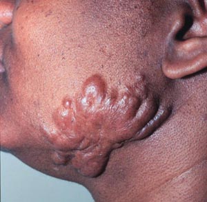
A keloid scar that developed from a skin wound along the edge of the jaw. The tendency to develop keloids is a genetic trait (Source: Leonard C. Sperling, MD.)
Hypertrophic Scars
Hypertrophic scars are caused by excess deposition of collagen fibers in a healing wound. This happens in burns, infected wounds, and wounds healing under tension. In hypertrophic scars, the excessive formation of collagen usually stops within a few weeks. The result is a scar that is thicker than normal and raised above the plane of the skin; but unlike a keloid, a hypertrophic scar does not expand out beyond the actual wound. Hypertrophic scars, which usually get smaller spontaneously, can occur anywhere on the body.
Hypertrophic scars are also produced in wounds that have a long reaction (inflammatory) healing phase and in which re-epithelialization has been delayed, such as in many burn wounds. For burn patients, continuous pressure (constant pressure lasting 6 to 12 months) can help to reshape and flatten hypertrophic scars. Specialized secondary pressure dressings are available for hypertrophic-susceptible and burned areas such as the face and hands.
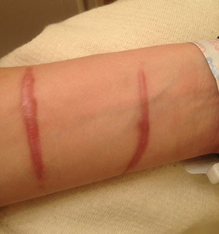
Hypertrophic scar, four months after incident. (Source: Cgomez447, CC BY-SA 3.0.)
Contractures
All scars go through a process of shrinking or contracting. Enlarged scars, however, sometimes contract excessively, with extreme tightening and constriction of the skin surface, leading to physical defects and functional disability (Johns Hopkins, 2022). When contractures form over joints, the scars can make bending difficult or impossible. Disabling contractures most commonly form across finger joints, along the neck, across the axilla, and across the antecubital fossa.
Contractures after amputation surgery of a lower extremity can occur in up to 5% of cases and can begin to develop within a few days after the surgery. Contractures usually occur in the joint nearest to the amputation site, such as the tibiofemoral joint or the acetabulofemoral joint.
A contracture is a permanent fixture of the skin, and it cannot be repaired by stretching, massaging, or applying ointments, lotions, or creams. The most successful treatment for a contracture is to have it excised surgically. Early consultation with physical and/or occupational therapy can be an important step in the prevention of contractures during the wound healing process.
Types of Wound Closure
Wound closure is described as:
- Primary closure (primary intention)
- Secondary closure (secondary intention)
- Tertiary closure (tertiary intention)
In primary closure the layers of involved tissue are brought together, and the wound edges are approximated and then surgically closed with either sutures or skin staples. Wounds closed by primary intention require only a limited amount of collagen to repair the tissue damage.
Secondary wound closure occurs when wounds are left open after surgery. An example of this is an abdominal wound repaired to the level of the fascia, with the remaining layers above this left open. These wounds fill in with new granulation tissue over a period of time, followed by wound contraction, and re-epithelialization. How long this takes depends on the overall condition of the patient and is affected by the presence of concomitant conditions such as cardiac disease and diabetes. Secondary wound closures tend to leave a larger scar. Other examples of wounds that heal by secondary intention are dehisced surgical wounds and pressure ulcers/injuries.
Tertiary intention, or closure, combines primary and secondary intention wound repair. The wound is allowed to fill in with granulation tissue and is then surgically closed. Delayed primary closure is used for highly contaminated wounds that may need repeated debridement or may need to be treated with antibiotics before being closed.
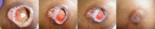
A pressure ulcer healing by secondary wound closure. Periosteum of bone is visible in the left picture. Healthy granulation tissue covers the wound in the two middle pictures. Healing took several months. (Source: Charlie Goldberg, MD, © Regents of the University of California.)
The main factors determining whether a wound will be closed immediately by primary intention or left open to heal by secondary or tertiary intention is whether there is a high risk of infection and whether the degree of tissue loss is such that the wound edges cannot be easily approximated without putting undue tension on the incision line. The immediate primary closure of a well-cleansed wound protects it from new contamination and allows the most control over the size and appearance of the final scar.
Wounds closed with sutures add new foci for infection (i.e., the suture holes), and sutures should not be left in place longer than is necessary. It is recommended that sutures be removed within one to two weeks after their placement. However, sutures should not be removed prematurely, since this may increase the risk of dehiscence and an increase in scar tissue. The following table lists optimal suture removal times for specific anatomic locations.
| Location | Time (days) |
|---|---|
| (Ratner, 2020) | |
| Face | 5–7 |
| Neck | 7 |
| Scalp | 10 |
| Arms and trunk | 10–14 |
| Lower extremities | 14–21 |
DEHISCED WOUNDS
When a wound that has already been closed spontaneously reopens, this is referred to as a dehisced wound. Incomplete dehiscence occurs when the skin edges separate but the deeper layers of tissue remain together. In complete dehiscence all layers of the wound separate, and this can extend down to and beyond the fascia. Evisceration happens when the intestine protrudes into the wound; it is a medical emergency (WOCN, 2022).