SKIN—THE EXTERNAL BARRIER
To properly understand the occurrence, continuation, and healing of wounds, it is necessary to first look at the skin.
The skin is comprised of two layers: the epidermis, made up of four to five thin layers stacked on top of each other, and the dermis (often referred to as the “true skin”), a layer of connective tissue directly below the epidermis. Beneath the dermis is the subcutaneous tissue, which separates the skin from the underlying muscles, tendons, joints, and bones. Skin varies in thickness from less than 1 millimeter in the eyelids to greater than 4 millimeters on the soles of the feet.

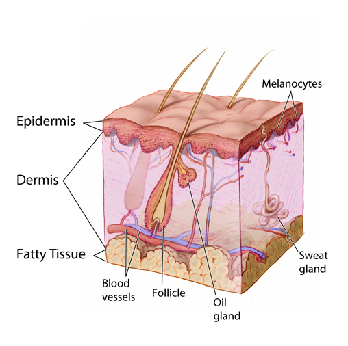
The layers of skin and associated glands and vessels (epidermis, dermis, fatty tissue, blood vessels, follicle, oil gland, sweat gland, and melanocytes). (Source: Don Bliss/National Cancer Institute.)
Epidermis
The deepest layer of the epidermis is known as the stratum basale or stratum germinativum. It is a layer of dividing, reproducing cells that migrate upward. As they travel, they differentiate and become filled with keratin, a tough, fibrous protein. The mature cells are pushed to the surface, where they die; thus, the outermost layer of the epidermis is made of flat, dead keratinocytes. The stratum basale also contains melanocytes, the cells responsible for producing melanin, the pigment that adds color to the skin and also protects against the damaging effects of ultraviolet light.
Shallow wounds to the epidermis usually heal rapidly and without complications. Usual skin thickness is reestablished, and there is no scar formation.
Dermis
The layer of skin directly beneath the epidermis is the dermis. A basement membrane separates these two layers. The dermis is mainly connective tissue and is therefore much stronger than the epidermis. The dermis varies in thickness across the surface of the body, but everywhere it is significantly thicker than the overlying epidermis.
The dermis is itself made up of two layers: the papillary layer is directly beneath the epidermis, and the reticular layer is below that. The primary function of the papillary dermis is to supply nutrients to the epidermis. The reticular dermis contains fibroblasts cells, which synthesize the connective tissue proteins, collagen, and elastin. These cells are responsible for the strength and elasticity of the skin. The reticular layer of the dermis also contains macrophages, which are essential for wound healing, and mast cells, a key component of the immune system.
The tissue of the dermis also contains small blood vessels, lymph vessels, nerves with their endings, and the smooth muscle fibers of hair follicles. Hair, nails, sweat glands, and sebaceous glands are sunken epidermal appendages that lie in deep valleys in the dermis surrounded by a row of germinative epidermal cells.
Subcutaneous Tissue
Beneath the dermis is a layer of subcutaneous tissue (also known as the hypodermis) containing fat. The thickness of the subcutaneous layer varies throughout the body. It is thickest along the anterior thigh and thinnest on the back of the hands.
Besides fat cells, subcutaneous tissue contains blood vessels, lymph vessels, and nerves. The subcutaneous layer is held together by a continuous sheet of fibrous membrane that runs parallel to the surface of the skin. This membrane is called the superficial fascia. The subcutaneous tissue provides insulation to the body, and it is also important in pressure redistribution (Baranoski & Ayello, 2020).
Gender differences are noted in the thickness and distribution of subcutaneous tissue. Males are inclined to have more subcutaneous tissue in the abdominal area and shoulders, while females have more subcutaneous tissue around their buttocks, hips, and thighs (Brannon, 2019).
Individuals with a thin layer of subcutaneous tissue and limited mobility are at high risk for the development of pressure ulcers/injuries. Those with excess subcutaneous tissue can have a harder time with wound healing since subcutaneous tissue is not well perfused, and this in turn decreases the blood supply to the dermis and epidermis (WOCN, 2022).
The subcutaneous tissue is a loosely organized compartment. When skin wounds extend deeper than the dermis, dirt is easily pushed into and spread within the subcutaneous tissue. This increases the risk of infection and requires that deep wounds be cleaned thoroughly.
Beneath the subcutaneous tissue layer, structures such as muscles and organs are enclosed in their own separate connective tissue sheaths. The generic name for these sheaths is deep fasciae. Deep fasciae generally look off-white in fresh wounds. When treating a wound, tears in the deep fasciae are repaired whenever possible.