NUCLEAR/RADIOLOGICAL TERRORISM PREPAREDNESS AND RESPONSE
Nuclear, or radiological, weapons are the newest member of the WMD “family.” So far, two nuclear weapons have been used, both by the United States near the end of World War II. These weapons ended the war sooner and resulted in the immediate deaths of approximately 120,000 people.
Nuclear terrorism might involve the acquisition of or the fabrication of a nuclear weapon such as a radiological dispersal device (dirty bomb). A dirty bomb is a mix of explosives, such as dynamite, with radioactive powder or pellets. When explosives are set off, the blast carries radioactive material into the surrounding area. Other radiation emergencies may involve an incident at a nuclear power plant, which could release radiation over an area.
Creating a nuclear WMD requires a high degree of scientific knowledge (or the ability to interpret and apply information available on the Internet) as well as access to specific materials and facilities. These technical challenges for making a nuclear explosive, however, should not be regarded as insurmountable. Unlike state-sponsored nuclear weapons developers, terrorists have different requirements for safety, performance, and delivery. Also, the rapid availability of technological knowledge can further advance terrorists’ weaponization attempts.
Despite thefts of small amounts of fissile material, however, there is no credible evidence that any terrorist group has succeeded in obtaining the necessary multi-kilogram critical mass of weapons-grade plutonium required to make a nuclear weapon (Williams et al., 2021).
NEVADA NATIONAL NUCLEAR SECURITY SITE
A national nuclear security site that uses and creates nuclear material is located just 60 miles from the Las Vegas urban area. Materials delivered to and from the site travel directly through the urban area, increasing the risk for a terrorist attack that could release radioactive materials into the community. To protect the community, agencies have developed a preventive radiological nuclear detection program designed to prevent the illicit development, transport, or use of radiological or nuclear materials anywhere in the Las Vegas urban area. This involves primary and advanced secondary radiological screeners and related detection equipment (FEMA, 2021).
What Is Ionizing Radiation?
Ionizing radiation has so much energy that it can knock electrons out of atoms, a process known as ionization. Ionizing radiation can affect the atoms in living things, so it poses a health risk by damaging genetic tissue and DNA. Sources of ionizing radiation include X-ray machines, cosmic particles from outer space, and radioactive elements such as plutonium. Radioactive elements emit ionizing radiation as their atoms undergo decay. This can be a positive attribute when it is used as a method of medical treatment or negative when it is harnessed into a weapon.
MEASURING RADIATION
There are different but interrelated units for measuring radioactivity and its effects. These include:
- Radioactivity: The amount of ionizing radiation released by a material, representing how many atoms in the material decay in a given time period. The units of measurement for radioactivity are the becquerel (Bq, international unit) and the curie (Ci, U.S. unit).
- Exposure: The amount of radiation traveling through the air. The units for exposure are the coulomb/kilogram (C/kg, international unit) and the roentgen (R, U.S. unit).
- Absorbed dose: The amount of radiation absorbed by an object or person. The unit for absorbed dose is the gray (Gy, international unit) or the rad (U.S. unit). One gray is equal to 100 rads.
- Effective dose: The amount of radiation absorbed by a person, adjusted to account for the types of radiation received and the effect on particular organs. The unit used for effective dose is sievert (Sv, international unit) or rem (U.S. unit).
(EPA, 2021)
Because radiation cannot be detected by human senses, a device must be used to confirm or exclude its presence. In the case of a radiation terrorism incident, two general types of devices will be used, one to survey victims or healthcare workers and the other to monitor healthcare workers’ cumulative exposure.
Radiation Survey Meters
Colloquially known as Geiger counters, radiation survey meters identify the presence of radiation in the physical environment or on the surface of or within victims. Geiger counters can detect the presence of radiation but cannot determine the original source of the radiation, what type it is, or how much energy it contains. They are used during triage and decontamination of victims.
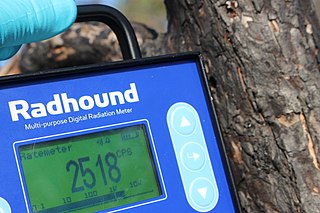
Radhound Geiger-Müller radiation detector. (Source: ArticCynda, 2019.)
Personal Dosimeters
A personal dosimeter detects and measures radiation that an individual has been exposed to. These devices detect high-energy beta, gamma, or X-ray radiation and are required for workers who operate X-ray machines, fluoroscopy units, and those working with radioisotopes or are exposed to other sources of radiation.
Whole-body dosimeters are badges or pocket devices that can be worn anywhere on the body, including the head and neck. Whole-body dosimeters are used in a wide range of occupational environments, including research, healthcare, and by emergency response providers.
Electronic whole-body dosimeters are worn anywhere on the body to detect and monitor exposure to X-ray, beta, and gamma radiation in real time. Radiation is detected and processed to give a real-time readout to the user for deep dose, skin dose, and dose rates. These dosimeters are used by pregnant workers and emergency response providers.
Extremity dosimeters come in ring, wrist, and ankle models to measure exposure in those individuals who are at risk for extremity exposure. These may include researchers using radioactive materials, individuals who use X-ray machines, individuals who work with radiopharmaceuticals, and individuals who may be required to hold patients during X-ray procedures (Government of Canada, 2020).
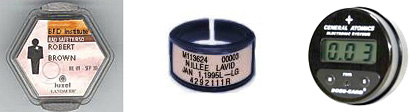
Dosimeters: film badge (left), ring badge (center), and real-time (right). (Sources: OSHA and U.S. DHS.)
Health Effects of Radiation
Exposure to radiation can cause two kinds of health effects:
- Deterministic effects are observable and can occur soon after receiving a large dose. These may include hair loss, skin burns, nausea, or death.
- Stochastic effects are long-term, such as cancer.
The radiation dose determines the severity of a deterministic effect and the probability of a stochastic effect. The goal of radiation control is to prevent any deterministic effects and minimize the risk for stochastic effects (CDC, 2021d).
TYPES OF RADIATION EXPOSURE
Radioactive contamination occurs when radioactive material is deposited on or in an object or a person. Radioactive materials released into the environment can cause air, water, surfaces, soil, plants, buildings, people, or animals to become contaminated. A person exposed to radiation is not necessarily contaminated. For a person to be contaminated, radioactive material must be on or inside the body.
There are four different types of injury that can be induced by radiation—irradiation, external and internal contamination, and incorporation of radioactive material.
- Irradiation or radiation exposure occurs when all or part of an individual’s body is exposed to penetrating radiation. An example of irradiation is the process involved with an ordinary chest X-ray. Following irradiation, the individual is not radioactive and cannot spread radioactive contamination.
- External contamination involves contamination with radioactive material, which can be in the form of dust, powder, or liquid, to the skin, hair, or clothing. External contamination requires removal of contaminated clothing and washing the material off.
- Internal contamination occurs when a person swallows or breathes in radiative materials or when radioactive materials enter the body through an open wound or by absorption through the skin. Some types of radioactive materials stay in the body and are deposited in different body organs, while other types are eliminated through blood, sweat, urine, and feces. Once the material has been eliminated from the body, the individual does not pose a risk to others.
- The fourth type of radiation injury involves incorporation or uptake of radioactive materials by body cells, tissues, and target organs such as bone, liver, thyroid, or kidney. Radiation materials are distributed throughout the body based upon their chemical properties.
(CDC, 2021d)
RADIATION ILLNESS
Once a person has been exposed to radiation beyond a therapeutic dose, radiation-induced illness may occur. Two major categories have been identified: acute radiation syndrome and cutaneous radiation syndrome.
Acute Radiation Syndrome (ARS)
Acute radiation syndrome is caused by total or near-total body irradiation by a high dose of external, penetrating radiation over a very short period of time (minutes). Onset and severity of symptoms are related to the severity of exposure. Victims in close proximity to the detonation of either a nuclear device or a “dirty bomb” would receive the intense exposure that causes ARS. The major cause of this syndrome is depletion of immature parenchymal stem cells in specific tissues (CDC, 2021d).
There are three classic acute radiation syndromes:
- Hematopoietic syndrome: Lymphocytes die from radiation-caused apoptosis, and precursor cells in the bone marrow are destroyed. This prevents production of leukocytes and platelets; gradually, circulating cells die and are not replaced. The syndrome progresses to infections and possible hemorrhage.
- Gastrointestinal syndrome: Irradiation causes mucosal stem cell death in the intestinal glands (colonic crypts), and new cells cannot be produced. This results in denudation of the gastrointestinal tract and the spread of bacteria. Death usually occurs in 3 to 10 days.
- Cardiovascular/central nervous system syndrome: Irradiation causes vascular damage resulting in significant cerebral edema and circulatory collapse. The patient develops nausea, vomiting, ataxia, hypotension, tachycardia, convulsions, and coma. No recovery is expected, and death usually occurs within 3 days of exposure.
(Williams et al., 2021)
Each of these syndromes go through four stages, as described in the table below.
| Stage | Characteristics |
|---|---|
| (CDC, 2021d) | |
| 1. Prodromal (early) |
|
| 2. Latent |
|
| 3. Manifest illness |
|
| 4. Recovery or death |
|
Acute Radiation Syndrome Treatment
Treatment for persons with internal exposure:
- Initial treatment includes lavage with fluids and charcoal to minimize absorption of radioactive materials.
- Radioactive iodine can be used with saturated solution of potassium iodide within a few hours to decrease the uptake of radionuclide in the thyroid. This may decrease the risk of malignancies in the future.
- Penicillamine is a chelating agent that binds to specific radioactive metals and results in decreased tissue uptake and increased excretion.
- Cesium exposure can be treated with ferric hexacyanoferrate, which will decrease gastrointestinal absorption.
- Treatment of exposure to americium, curium, and plutonium can include Ca-DTPA and Zn-DTPA.
Treatment for individuals with large-dose radiation exposure includes:
- Fluids and electrolytes are administered for managing nausea, vomiting, and diarrhea.
- Depending on the dose of radiation, antibiotics, cytokines, transfusions, and platelet transfusion should be considered.
- If absolute neutrophil count is <500 cells/mm, prophylactic antibiotics and antiviral, antifungal, and antipseudomonal coverage are considered.
- Filgrastim may be considered for treatment of hematopoietic syndrome.
(Williams et al., 2021)
Cutaneous Radiation Syndrome (CRS)
CRS can occur without symptoms of ARS. This is especially true with acute exposures to beta radiation or low-energy X-rays, because these are less penetrating and less likely to damage internal organs than gamma radiation. Most cases of radiation-caused skin injury have occurred when people inadvertently came in contact with unsecured radiation sources. In addition, cases of CRS have occurred in people who receive radiation therapy for cancer.
Exposure to radiation can damage the basal cell layer of the skin. Early signs and symptoms of CRS inflammation include itching, tingling, and dry or moist desquamation. In addition, radiation damage to hair follicles can cause epilation.
Transient and inconsistent erythema (associated with itching) can occur within a few hours of exposure and be followed by a latent, symptom-free phase lasting from a few days to several weeks. After the latent phase, intense reddening, blistering, and ulceration of the irradiated site are visible. Depending on the radiation dose, a third and even fourth wave of erythema are possible over the ensuing months or possibly years.
In most cases, healing occurs by regenerative means; however, large radiation doses to the skin can cause permanent hair loss, damaged sebaceous and sweat glands, atrophy, fibrosis, decreased or increased skin pigmentation, and ulceration or necrosis of exposed tissue.
Cutaneous radiation burns should be treated similarly to thermal burns. Severe burns may require amputation, grafts, or vasodilator therapy (CDC, 2021d).
Management of Response to a Nuclear/Radiation Terror Attack
The initial care team should include staff with expertise in radiation safety as well as trauma-related injuries. The facility disaster plan should contain information on how to contact individuals with this type of experience. For facility staff, an up-to-date alert roster should be readily available. If this expertise is not available in-house, a consultative relationship with a larger institution may be obtained. This should be included in the disaster plan.
In the absence of either of these options, or to augment them, the Radiation Emergency Assistance Center is available 24/7/365 (see “Resources” at the end of the course). This agency’s function includes deploying to and providing emergency medical consultation for incidents involving radiation anywhere in the world.
HOSPITAL TRIAGE
An ad hoc triage area should be established on location based upon a preestablished disaster plan and anticipated number of casualties, with a contaminated area and clean area separated by a buffer zone. Whenever possible, all triage participants should be volunteers. Healthcare providers should work in teams and a radiation safety officer should monitor them for exposure with dosimetry.
When a facility is expecting only one or two patients with possible radiologic contamination, triage can be done in the ambulance or ambulance bay. Moving triage outside of the facility during a mass casualty incident helps to prevent large numbers of patients from presenting directly to the ED.
The triage area should allow for rapid setup and have adequate staffing, supplies, and radiation monitoring capability to sort patients by medical severity. “DIME” is the standard NATO nomenclature recommended for triage:
- Delayed: May be life-threatening, but intervention can be delayed
- Immediate: Immediate attention required to prevent death
- Minimal: Ambulatory, minor injuries, can wait for definitive attention
- Expectant: Survival unlikely
Those patients with immediate life-threatening injuries should be brought directly to the emergency department prior to radiologic survey and decontamination. For stable patients with serious illness or injury, radiologic decontamination should be done prior to the patient being taken into the clean area of the emergency department.
Whenever possible, patients who are stable but contaminated with urgent injuries should enter the facility through a separate entrance and be taken to a designated decontamination room separate from the rest of the ED. If a separate entrance is not available, the areas where contaminated patients walk should be taped off and labeled as radioactive (Allen et al., 2021; Hamm, 2020).
STAFF PROTECTION GUIDELINES
Basic protective actions involve three factors: time, distance, and shielding. Radiation doses should be kept to a level that is as low as reasonably achievable. This is accomplished by:
- Minimizing time spent in the area of radiation; planning emergency response missions efficiently so that first responders enter and leave areas where they may be exposed or become contaminated as few times as possible and spend as little time as possible in the area
- Maximizing the distance between worker and source of radiation exposure; performing only life-saving and other critical tasks near a dangerous radiation source
- Using proper hazard controls, including shielding workers from a radiation source and contamination and the use of personal protective equipment
- Reassigning pregnant workers to job duties that minimize radiation exposure
- Conducting hazard assessment for workers and monitoring workers’ radiation doses
- Establishing an on-scene decontamination facility
(OSHA, n.d.-b)
PERSONAL PROTECTIVE EQUIPMENT
In a radiation emergency, choice of personal protective equipment depends on the response role, specific tasks, and risk of contamination. It is important to be aware that there is no practical personal protective equipment that can protect responders from high energy, highly penetrating forms of ionizing gamma radiation associated with most radiation emergencies.
Emergency responders arriving at a radiation emergency scene may not know they are being exposed to radiation unless they utilize a radiation detecting device. Monitoring devices are the only means to make certain that responders do not enter an area where exposure is excessive.
Level B PPE is used by first responders to prevent skin contamination by alpha and beta particles. Typical firefighter gear is considered adequate. Protection of internal organs from radioactive materials may be provided by wearing an appropriate particulate respirator. Protection against internal organ contamination can be provided by wearing an appropriate particulate respirator. SCBAs provide a high level of protections. Responders should use a full-face air-purifying respirator with a P-100 or HEPA filter.
Level C PPE is recommended for first receivers who are caring for victims highly suspected to be contaminated with radiological material. This includes a hooded, NIOSH-certified powered air-purifying respirator including appropriate breathing filters. A non-powered air-purifying respiratory may be worn when data confirm that a negative pressure respirator will adequately protect users from identified inhalation hazards.
Several types of PPE for personnel providing care for radiologically contaminated patients consists of modified universal precautions. Typically, these include:
- Shoe covers
- Zip-up coveralls (waterproof)
- Surgical cap
- Respiratory masks
- Face shield
- Inner pair of gloves
- Outer pair of gloves (different color than inner pair)
- Tape to secure outer coverings at junction of first pair of gloves with sleeves and show covers with coverall pants legs
- Personal radiation dosimeter worn so that a worker can easily see the read-out and/or hear warning alarms
(Williams et al., 2021; U.S. DHHS, 2021c; Allen et al., 2021)
Decontamination
Unless they have undergone a radiological survey and decontamination prior to arrival at the receiving hospital, all patients are presumed to be contaminated. The patient is surveyed with a Geiger-Mueller probe for external contamination as well as for fragments of radioactive shrapnel embedded in wounds. Radioactive fragments are promptly removed with forceps and sealed in lead containers. Findings are documented on an anatomic chart.
PERFORMING A SURVEY FOR RADIATION CONTAMINATION
In the event of suspected radiation contamination, healthcare personnel use a radiation survey monitor to survey patients.
Prior to beginning the survey:
- Inspect the equipment.
- Perform a battery check.
- Conduct a source/operational check.
- Conduct a background reading.
When performing the survey:
- Start at the head on the front side of the body.
- Continue systematically over the body, including the feet and soles.
- Repeat on the back side of the body.
(Source: Radiation Emergency Assistance Center/Training Site.)
(U.S. DHHS, 2021d)
Decontamination includes removal of clothing and washing. This process is capable of removing 90%–95% of external contamination.
- Remove patient clothing carefully to avoid spread of contamination. Double-bag clothing per radioactive hazardous waste guidelines, label, and save as evidence.
- Cleanse contaminated area. Wash first with saline. If there is facial contamination, flush eyes, nose, and ears with saline and rinse the mouth.
- Cleanse skin with soap and water, beginning with the areas of highest contamination.
- Resurvey and note radiation levels.
- Repeat washing until survey indicates radiation level is no more than 2–3 times the background level or the level remains unchanged.
- Cover wounds with waterproof dressing.
- Dispose of waste water through normal channels.
(Allen et al., 2021)
ADDRESSING INTERNAL CONTAMINATION
Internal contamination is considered if high survey readings persist following decontamination.
Nose or mouth contamination may indicate inhalation or ingestion. The patient is scanned and each nostril is swabbed separately to help estimate the level of internal lung contamination. A spot urine sample is also obtained for isotope measurement.
Internal contamination continues until the radioactive material decays, is flushed from the body by natural processes, or is removed by medical countermeasures. Decision to treat will depend on:
- Level of internal contamination
- Size of radiation event
- Availability of resources/personnel
- Likelihood the patient will survive
(U.S. DHHS, 2021e)
CASE
A “dirty bomb” explodes in a crowded casino. Per local emergency response plans, all hospitals in the area are notified to expect the arrival of multiple casualties with both radiation exposure and traumatic injuries. ED staff and others involved in each facility’s emergency response team don appropriate PPE prior to the arrival of the first victims.
Per protocols, treatment is begun immediately for physiologically unstable patients prior to performing the initial survey or beginning initial decontamination. For those without life-threatening injuries, initial radiation surveys are performed. Care providers then remove contaminated clothing from the individuals and place each victim’s clothing and belongings in individual property bags that are then properly labeled. Once all clothing is removed and secured, the secondary survey is performed.
In victims who have been injured by shrapnel from the blast, ED staff remove the shrapnel to protect against possible internal contamination. They clean the wounds, carefully catching any water run-off. They also wash the patients’ bodies with soap and water to remove any external contamination. They then complete a second scan to determine the degree to which the decontamination process was successful.
Psychosocial Aspects of Radiation Terrorism
Major contributing elements to psychosocial stress involved in a nuclear or radiological incident include the unknown nature of radiation and uncertainty related to the extent of risk for one’s health, the implementation of the protective actions, and the stigmatization of affected people.
Many of the psychosocial aspects of radiation emergencies are similar to those in other emergency situations. However, acute fear, psychological responses to somatic illnesses or injuries, and long-term development of medically unexplained symptoms are particularly likely in radiological or nuclear emergencies. A range of psychosocial concerns are therefore taken into account when planning for radiation emergencies.
Individuals who may require mental and psychosocial support may include:
- People in close proximity to the radiological event
- First responders, healthcare personnel, clean-up workers, and other responders working under hazardous or stressful conditions
- Parents and future parents concerned about long-term genetic effects of radiation
- Evacuees and members of hosting communities
- Persons with pre-existing mental health and psychosocial needs
- People with low literacy, who may struggle to follow advice and instructions provided by risk communicators
(WHO, 2020)
COMMUNICATION
Communication is vital in order to inform people of the health risks they face. Accurate information provided early, often, and in languages that the people understand and through channels people trust enables them to make choices and take actions to protect themselves.
Emergency risk communication is an important aspect of the response to a nuclear incident to help mitigate stressors. This involves the real-time exchange of information, advice, and opinions between experts, community leaders or officials and the people who are at risk (WHO, 2020).
Reporting a Radiation Incident
To report an incident involving nuclear materials, contact the federal government’s centralized reporting center. The U.S. Nuclear Regulatory Commission should be notified of:
- Any accident involving a nuclear reactor, nuclear fuel facility, or radioactive materials
- Lost or damaged radioactive materials
- Any threat, theft, smuggling, vandalism, or terrorist activity involving a nuclear facility or radioactive materials
For guidance in the management of a nuclear emergency, the Radiation Emergency Assistance Center (REAC/TS) should be contacted to record the incident and to receive expert guidance on the medical management of radiation incidents. REAC/TS provides emergency response and subject matter expertise on the medical management of radiation incidents for the National Nuclear Security Administration’s (NNSA) Office of Counterterrorism and Counterproliferation.
(See also “Resources” at the end of this course.)