HEALING ASSESSMENT AND DOCUMENTATION
Pressure injuries are assessed initially and reassessed at least weekly, with careful documentation of the findings. Minimally, wound care assessment and documentation include:
- Wound measurements (length, width, depth)
- Presence or absence of tunneling and undermining and its exact location (e.g., “undermining present from 9 o’clock to 11 o’clockto a depth of three centimeters”)
- Amount of drainage present (large, moderate, scant), its color, and presence of an odor (e.g., “a moderate amount of serosangenous drainage, no odor noted”)
- Condition of the periwound area (e.g., “periwound tissue is dry and intact with a small 2-cm area of redness at the distal end of the wound)
- Patient’s response to any dressing change
- Whether pain medication was administered prior to the procedure
With each dressing change, the injury is also observed for anything that may indicate the need for a change in treatment (e.g., improvement or deterioration, more or less drainage, signs of infection, or other complications). Any signs of deterioration should be addressed immediately. The type of dressing may need to be changed based on this assessment (e.g., to an antimicrobial dressing or a more absorptive dressing) and/or a change made in frequency of wound care.
Signs and Factors in Wound Healing
General signs of healing are decreased size, less exudate, and tissue changes from devitalized tissues (slough and eschar) to granulation tissue and epithelialization.
- Stage 1 and 2 pressure injuries should show evidence of healing within one to two weeks.
- Stage 3 and 4 pressure injuries should show evidence of healing within two to four weeks.
- Large, deep, infected pressure injuries and those with large amounts of drainage and/or covered with slough or eschar are significantly less likely to heal within even three months, and some may not be fully healed even after five or six months of treatment.
(WOCN, 2016a, EPUAP/NPIAP/PPPIA, 2019)
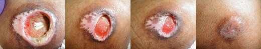
A healing pressure injury. Periosteum of bone is visible in the left picture. Healthy granulation tissue covers the wound in the two middle pictures. Healing took several months. (Source: Charlie Goldberg, MD, © Regents of the University of California.)
If after two weeks of treatment there has been no healing or signs of improvement, then all the risk factors are reevaluated and the plan of care revised to reflect new interventions.
In the case of a nonhealing pressure injury—and after the choice of wound care has been evaluated as appropriate and pressure is being relieved—then the patient is reassessed for other reasons why the injury is not improving. One systematic approach to determining what other factors might be affecting wound healing utilizes the acronym DIDN’T HEAL. Using this acronym and correcting those factors that can be corrected will aid in healing the injury. If factors cannot be corrected, healing the injury may not be possible.
| Cause | Description | Additional Factors | |
|---|---|---|---|
| (Daley, 2018) | |||
| D | Diabetes | Lack of diabetic control, causing diminished cardiac output, poor peripheral perfusion, and a decrease in the ability of WBCs to function |
|
| I | Infection | Increased destruction of collagen needed for repair |
|
| D | Drugs | Possible impaired collagen synthesis |
|
| N | Nutrition | Deficiencies impairing normal wound healing |
|
| T | Tissue necrosis | Lack of oxygen impairing healing |
|
| H | Hypoxia | Inadequate tissue oxygenation impairing healing |
|
| E | Excessive tension | Tension on wound edges leading to local tissue ischemia and necrosis |
|
| A | Another wound | Competition for factors needed for wound healing, impairing wound healing at all sites |
|
| L | Low temperature | Deceases to oxygen to the wound |
|
CASE
James, a 29-year-old male patient who is paraplegic, arrived to the emergency department because of a recurrent stage 4 pressure injury at his left ischium. The patient states that his pressure injury returned because his wheelchair support cushion malfunctioned and the resulting pressure reopened the injury at the site of the scar tissue. James underwent surgery to the same area five years ago, which promoted healing of the previous injury through removal of infected bone and a flap-graft surgical reconstruction.
The size of the reopened wound is extensive, and the amount of drainage saturates a large, thick dressing in 12 hours. There is no necrotic tissue, but the patient’s bone is visible. Due to sepsis, James is admitted to the hospital. The infectious disease physician prescribes IV antibiotics for probable osteomyelitis. A surgical consult suggests possible surgery to the area again to close the wound, but not until the infection is cleared and the patient’s nutritional status is optimized.
To manage the drainage and protect the wound, a negative-pressure wound therapy dressing system is applied per protocol. The dietitian assesses James and recommends improvements to his nutritional status, particularly his protein intake. After a week in the hospital, James is discharged with home healthcare for six weeks of IV antibiotics and negative-pressure wound therapy. Referrals are made to the social worker for resources, the physical therapist for an appropriate wheelchair cushion recommendation and functional mobility evaluation, the occupational therapist to assess the patient’s self-care skills at home, and the dietitian for continued recommendations. A low-air-loss mattress is obtained to reduce pressure while he is in bed.
The physical therapist visits James at home to evaluate his mobility. The patient is able to move himself in bed and to safely transfer himself from the bed to a chair.
The occupational therapist also visits James at home, completes an evaluation, and identifies that he is independent with his activities of daily living. The occupational therapist observes James’s abilities for meal preparation and suggests modified utensils and a long-handled reacher that will make it easier for James to prepare meals.
The home health nurse visits James three times per week to change his wound dressing, draw labs, change the IV dressing, and assess his adherence to self-administering the IV antibiotics. The home health dietitian finds a source for an affordable protein supplement for James’s loss of protein from his wound drainage.
Healing Assessment Tools
There are several tools for assessing pressure injury healing. The Bates-Jensen Wound Assessment Tool (BWAT) is comprised of 15 items, of which 13 are scored from 1 to 5. The total scores and dates of assessment can be plotted on a graph, which provides an index of improvement or deterioration of the wound.
The PUSH tool (Pressure Ulcer Scale for Healing) was developed by NPIAP. A pressure injury is categorized using numerical scores of 0 to 5 according to surface area (length multiplied by width), drainage amount, and tissue type. A comparison of the total scores measured over time provides an indication of improvement or deterioration in the injury.
The Spinal Cord Impairment Pressure Ulcer Monitoring Tool (SCI-PUMT) was developed to assess pressure injury healing in patients with spinal cord injury. Pressure injury healing is defined as the reduction in volume of the pressure injury and complete healing as resurfacing of the wound. Volume is an estimate obtained by multiplying length by width by depth (WOCN, 2016a).
Many computer systems also have programs to monitor pressure injury progress. Of course, the clinician will also use clinical judgment to assess signs of healing, such as a decrease in the amount of drainage, pain, and wound size and an improvement in wound bed tissue. The clinician can also use photography, comparing baseline and serial photographs to monitor healing over time. Follow facility policy on the use of photography (see “Photography” below).
(See also “Resources” at the end of this course.)
Documenting the Healing Process
Wound care documentation includes a variety of information that reflects the wound status while it heals. Providing an accurate description of the skin and wound characteristics is critical following each dressing change. These findings of the pressure injury’s current status will help the clinician in revising the plan of care and treatment strategies over time.
The very basics of documentation are to document what was observed, what was done (including education provided), and how the patient responded. Documentation of pressure injury management includes an assessment of the pressure injury on admission, with each dressing change, on transfer, at discharge, or when a change in condition occurs (and per agency regulations) for any signs of skin and/or wound improvement or deterioration. Documentation also includes risk assessment and patient/family education provided. The skin under and around medical devices in particular is assessed for injury twice a day or more often if patient is prone to edema.
The following parameters are documented:
- At admission, onset, course, and duration of the pressure injury
- Description of the pressure injury
- Pain (location, intensity, quality, onset, duration, alleviating/aggravating factors)
- Patient/caregiver’s ability and willingness to adhere to the prevention and treatment program
- Prevention interventions that were initiated (referrals to dietary, physical therapy, occupational therapy, support surface management, skin care management, etc.)
- Discussions conducted with and observations made by physicians
The following elements are documented in any wound assessment (WOCN, 2016b; EPUAP/NPIAP/PPPIA, 2019):
ANATOMIC LOCATION
The anatomic location of the pressure injury is identified using proper terminology in documentation. (Terms such as anterior-posterior, medial-lateral, or proximal-distal can clarify location.) Anatomical drawings or photography may be used.
STAGING
The stage of the pressure injury is determined and documented. (See “Staging Pressure Injuries” earlier in this course.)
DRAINAGE/EXUDATE
Color, type, consistency, and amount are identified and documented. Amount may be indicated as: none, light/scant, moderate, heavy/large, or copious. Color may be indicated as serous (clear, watery plasma); sanguineous (bloody); serosanguineous (plasma and red blood); or purulent (thick, odorous, possibly yellow, green, or brown).
ODOR
Odor defines the presence or absence of high bacteria counts in the pressure injury and should be assessed only after cleaning the wound. Almost all drainage has an odor. A strong or foul odor from the wound bed suggests infection. A mild odor may be due to the particular wound care products in use.
DESCRIPTION OF WOUND EDGES
Wound edges may be documented as:
- Attached: Edges are attached, moist, and flush with the wound base (see wound images earlier in this course).
- Unattached/rolled: Undermining is present between the dermis and subcutaneous tissues. The edge of the wound is raised and a lighter color than the surrounding tissue.
- Undermined: There is a gap in the edge of the tissue that creates a lip or overhang of the edge.
DESCRIPTION OF THE PERIWOUND SKIN
Periwound skin is observed at least 4 cm around the wound. The periwound skin should be intact. It is documented as to the following qualities:
- Color: There may be redness, pallor, blanchable erythema, nonblanchable erythema, or purple discoloration.
- Temperature: Warmth may indicate further tissue breakdown or underlying infection.
- Induration: Abnormal firmness with a definite margin may indicate infection.
- Maceration: Softening of tissues may be due to soaking from wound drainage or contact with urine and/or stool.
- Denuded: Superficial skin loss may be due to drainage or trauma (such as from tape). Excoriation refers to linear, scratch-like marks, not to skin loss from trauma or incontinence.
TYPE OF TISSUE EXPOSED (APPEARANCE OF WOUND BED)
- Red: This may indicate clean, healthy granulation tissue. Granulation is a pink or red moist tissue composed of new blood vessels and connective tissue that fills an open wound when it starts to heal. It usually has an irregular, granular surface, like velvet. Not all red tissue is granulation.
- Yellow: This may indicate the presence of drainage or slough. Slough is a soft, moist, avascular (lacking blood supply) tissue that may be yellow, white, tan, or green. It may be loosely or firmly attached. It sometimes resembles chicken fat. Not all yellow tissue is avascular; it could be fibrous.
- Black: This may indicate the presence of eschar or necrotic tissue, which slows healing and allows bacteria to grow. It may be brown or tan and can be hard or soft or loosely or firmly attached. It can resemble a scab, but there is no healing occurring under it.
(See “Staging Pressure Injuries” earlier in this course for images of types of exposed tissue.)
WOUND MEASUREMENTS
Measurements are done at least weekly and following debridement. It is not necessary to document measurements with each dressing change, as changes in wound size do not occur that rapidly.
Measurements are taken using a single-use, metric tape measure; “coins” (dime-sized, quarter-sized, etc.) are not used. Descriptors such as round, oval, irregular, etc., are also useful. If possible, the patient is in the same position each time the wound is measured to promote consistency of measurements. Communication of the wound size is useful for other members of the healthcare team, regulatory agencies, and payers to determine progress.
- Length: Linear distances are taken from wound edge to wound edge and measured consistently over time. One method is to look at the wound as if it were a clock face: the top of the wound (12 o’clock) is toward the patient’s head. The bottom of the wound (6 o’clock) is toward the patient’s feet. Length is the longest distance measured from head to toe, or 12 o’clock to 6 o’clock.
- Width: Width is longest distance measured from side to side, perpendicular to the length, or from 9 o’clock to 3 o’clock.
- Depth: This is the distance from the visible surface to the deepest point in the wound base. Depth can be measured using a cotton-tip applicator, holding it perpendicular to the wound edge, placing the finger at the point on the swab that corresponds to the wound edge. The swab is then removed, with the distance on the swab measured on the tape measure.
- Undermining: A cotton-tip applicator is used to probe to the deepest part of the undermining, marking the depth between the end of the applicator and the wound edge with the finger and measuring it against the tape measure. The location of the undermining can be indicated using the clock face (e.g., “undermining extends from 12 o’clock to 5 o’clock and is deepest at 3 o’clock at 3 cm”).
- Tunneling or sinus tract: The tract is measured as for undermining and its location described using the clock-face method.
PHOTOGRAPHY
Some healthcare facilities use baseline and serial photographs as a method of monitoring pressure injury progress over time and evaluating the effectiveness of the current wound care. Photographs do not replace bedside assessment but may be useful for documentation. Techniques and equipment must be standardized and staff trained in taking the types of photographs required to ensure an accurate representation of the condition of the pressure injury so it can be reliably compared over time (Ayello & Baranoski, 2016).
The frequency of photos depends on individual facility policies, but at a minimum, photos are taken the first time the wound is assessed, once healing has occurred, and when the patient is transferred to another care setting. Some facility policies require weekly wound photography.
Prior patient consent is required for wound photography, and the use and confidentiality of the photos must be thoroughly explained to the patient. A written facility policy on wound photography will address:
- Patient consent (including a form)
- Frequency of photography
- Staff authorized to take wound photos
- Methods of identifying the patient, for example, placing the patient’s initials, medical record number, date, and time on a measuring guide placed proximal to the wound and included in the photo
- Storage of the photos in the patient’s records and who will have access to them
(Nix, 2016; EPUAP/NPIAP/PPPIA, 2019)
CASE
After a few weeks of appropriate treatment, Mrs. Olivera, a patient with a pressure injury, remains in the hospital. The nurse manager reviews the nursing documentation, specifically the patient’s weekly wound measurements, for evidence that the wound is healing. The nurse manager detects a large variance between the patient’s wound measurements. On admission, Mrs. Olivera’s wound measured 4 cm x 6 cm x 3 cm (length x width x depth). A week later, the patient’s wound was documented to measure 1.5 x 2.5 x 1, indicating the wound had decreased in size dramatically. The third week the wound was documented as 5.5 x 3.5 x 2.5, indicating that the wound had worsened dramatically.
Such changes don’t make sense to the nurse manager. In questioning the staff about these measurement differences, the nurse manager discovers that for the second week’s measurements, the nurse reversed the measuring device and measured in inches rather than centimeters. For the third week, another nurse documented the width as the length and the length as the width.
At the next staff meeting, the nurse manager brings a wound model so that the nurses can practice wound measurement. The manager reviews that length is a head-to-toe or 12 o’clock–to–6 o’clock measurement, and that width is a side-to-side or 9 o’clock–to–3 o’clock measurement. She also emphasizes the need to use metric measurements. As a result, Mrs. Olivera’s wound is now consistently measured, demonstrating that the facility’s care of the patient is helping to heal the wound.
POTENTIAL COMPLICATIONS ASSOCIATED WITH PRESSURE INJURIES
- Heterotopic bone formation (presence of bone in soft tissue where bone does not normally exist)
- Fistula
- Abscess
- Osteomyelitis
- Bacteremia/sepsis
- Cellulitis
- Squamous cell carcinoma (Marjolin’s ulcer, an chronic ulcer that undergoes malignant transformation)
- Significantly higher risk for postoperative septicemia, pneumonia, stroke, urinary tract infection, and acute renal failure
- Higher risk of postoperative mortality in patients who have a pressure injury preoperatively
Minimizing the Recurrence of Ulcers
Achieving a closed wound is just the beginning of the effort to prevent a pressure injury from recurring. Clinicians must emphasize and reemphasize to patients and caregivers that measures to promote healing and prevent recurrence are lifelong. Recurrence rates for adults have been reported as high as 56%, and 21% develop a new injury at a different site (WOCN, 2016a). Patients with spinal cord injury have a pressure injury rate of 17% to 33% but have a recurrence rate from 31% to 79%.
The most common factors associated with recurrence are related to a lack of compliance with offloading the pressure area and maintaining a healthy lifestyle, such as stopping smoking, maintaining a normal weight, and controlling blood sugars if diabetic. Psychosocial problems (e.g., unemployment, low level of education, drug or alcohol abuse) have also been reported to increase the risk for pressure injury recurrence.
Clinicians must initiate and continue preventive education wherever an at-risk patient enters the healthcare system using interactive, individualized patient education. Telemedicine technology can be used to assess and teach patients who cannot easily come to a clinic or office.
Clinicians teach the following preventive measures to patients and caregivers:
- Perform a regular inspection of the skin, especially over bony prominences, using a mirror or even cell phones or digital cameras if necessary, to identify signs of pressure as evidenced by changes in the skin:
- Color, such as a reddish or purplish hue
- Temperature (warmer or cooler) compared to the surrounding skin
- Texture, such as bogginess or induration
- If skin changes are present, offload pressure to the area and recheck in 15 minutes; continue to monitor the skin until the skin change resolves, and notify a healthcare professional if it does not resolve.
- Follow appropriate skin care regimens:
- Keep the skin clean and dry.
- Use a mild soap and warm (not hot) water.
- Apply skin moisturizers such as petrolatum after bathing and when the skin is dry.
- For bed- and chair-bound patients:
- Use measures to reduce friction/shearing, such as lifting instead of dragging across the bed, and/or wearing clothing such as long-sleeved pajamas and socks.
- Routinely turn, reposition, and use pressure-redistributing devices if confined to a bed and/or chair.
- Avoid the use of rings, foam cut outs, or donut-type devices.
- Maintain adequate nutrition and fluid intake; monitor for weight loss, poor appetite, or gastrointestinal changes that interfere with eating; and promptly report changes in health and nutritional problems to healthcare providers.
(WOCN, 2016a; EPUAP/NPIAP/PPPIA, 2019)