PRESSURE INJURY TREATMENT
Treating a pressure injury involves all of the activities used in preventing a pressure injury: the proper pressure-reducing surface, repositioning the patient correctly and frequently, maintaining intact skin, and improving nutrition. While these interventions are started, the treatment of the wound itself also begins. There are basic wound care principles that can be used in deciding which treatments will be the best for the wound and for the patient. Frequent reassessment of the wound and its response to the treatment is required, as well as eliminating or reducing the factors that inhibit wound healing.
Of all the interventions that must be done to heal the injury, pressure reduction measures are the most important. Simply put, the wound will not heal unless the pressure is removed. Trying to heal a pressure injury without reducing the pressure is like trying to heal a stab wound with the knife still in it. There might be some improvement, but the wound will never heal because the primary cause has not been removed. (Techniques to reduce pressure are discussed earlier in this course.)
The object of pressure injury treatment is to reproduce (to the best of one’s ability) the normal environment of the exposed tissue of the wound. The normal environment of all tissue and cells, with the exception of the epidermis, is warm, dark, moist, and protected.
In order to heal any wound, including pressure injuries, some basic principles and management strategies need to be followed. The strategies for pressure injury treatment are:
- Cleanse the wound and periwound at each dressing change.
- Manage wound infections.
- Debride the pressure ulcer of devitalized or necrotic tissue.
- Utilize appropriate dressings.
Some of these strategies will require medical intervention; others, good clinical care. By carrying out these strategies, caregivers will provide the wound with the environment it needs to heal (WOCN, 2016a; EPUAP/NPIAP/PPPIA, 2019).
Pain Management
Before treatment begins, clinicians address the patient’s pain. Pressure injuries hurt. The pain may be constant and severe, and it may be the most distressing pressure injury symptom that the patient reports. Pressure injury pain can be caused by:
- Pressure, friction, shear
- Damaged nerve endings
- Inflammation
- Infection
- Procedures and treatments
(Bryant & Nix, 2016)
Multiple studies have shown that the pain increases as the stage increases (WOCN, 2016a). The words most frequently used to describe pressure injury pain in all stages (2–4) include “tender,” “hurting,” “burning/hot burning,” “sharp,” “throbbing,” and “aching.” The pain can occur when the patient is at rest and when no treatments are being done. The greatest pain occurs with dressing changes and wound care. Thus, pain must be assessed before, during, and after wound care and prevented/treated accordingly.
It should not be assumed that because the patient is paralyzed that the body cannot feel or respond to pain. Similarly, it should not be assumed that the patient with dementia, who never complains of pain, does not in fact have pain. The clinician should assume that if the wound would cause oneself pain, it is causing the patient pain. Assess and treat accordingly.
Some wound care strategies that can help reduce pain are:
- Organize care to coordinate it with pain medication administration.
- Encourage patients to request a “time out” during procedures, and then be sure to give a time out.
- Reduce the pain by keeping the wound bed covered and moist and using a nonadherent dressing.
- Select dressings that require less frequent changes such as hydrocolloids, hydrogels, alginates, foams, and soft silicone dressings.
- Protect the periwound skin with liquid film barriers or barrier ointments or creams to prevent skin damage from drainage and tape stripping.
- Encourage repositioning as a way to reduce pain, and use appropriate support surfaces.
- Provide pain medication prior to procedures.
(Bryant & Nix, 2016; EPUAP/NPIAP/PPPIA, 2019)
Topical analgesics can be applied to the wound prior to dressing changes. Topical lidocaine gels, creams, sprays, and patches are now available over the counter in concentrations as high as 5%. Lidocaine should be used with caution in wounds with large surface areas, as it can be absorbed through the wound and possibly cause neurologic or cardiovascular adverse side effects. EMLA cream (a eutectic mixture of local anesthetics, namely lidocaine and prilocaine) is another product used for topical analgesia. It must be in contact with the wound for at least 20 minutes under occlusion (e.g., plastic wrap over the top) and is quite effective (WOCN, 2016b).
CASE
Bill is a 76-year-old male with a history of multiple strokes (CVAs) and respiratory failure. He has a tracheostomy (but is able to breathe on his own), a G-tube for enteral feeding, and a urinary catheter. He’s been admitted with pneumonia and has community-acquired stage 4 pressure injuries on his sacrum and left ischium.
Bill cannot speak but makes eye contact, shakes his head yes and no appropriately to questions, and is usually smiling. He always shakes his head no when asked if he is having pain. After a few days of doing his wound care, the nurse notices that during care he is frowning, his breathing rate is a little faster, and he closes his eyes. When asked if he is having pain, Bill indicates no, but the nurse believes that he might be hurting.
The nurse discusses this with the physician, who agrees that a dose of oral pain medication can be given through the G-tube an hour before wound care. The physician also agrees to the nurse’s suggestion for the use of 2% lidocaine gel applied to the wound beds and allowed to remain in place for 10 to 15 minutes prior to wound care. With these additions to his care plan, Bill no longer frowns and seems a bit more relaxed during wound care. Bill’s son has noticed this as well and thanks the nurse for his father’s care.
Wound Cleansing
Cleansing is the important first step in preparing the pressure injury bed to heal by removing surface debris and dressing remnants and allowing better wound visualization for assessment. The goal is to flush away exudate without damaging tissues.
How often a pressure injury is cleansed is determined by the amount of drainage (e.g., heavily draining wounds may need to be cleansed three to four times a day), dressings used, and wound care treatment orders. The wound and periwound are cleansed at each dressing change, minimizing trauma to the wound.
While no specific studies demonstrate the superiority of a particular wound-cleansing product or technique for pressure injuries, the WOCN (2016a) offers the following recommendations:
CLEANSING SOLUTIONS
Most pressure injuries can be cleansed with potable water (i.e., water suitable for drinking), water that has been boiled and cooled, or normal saline. A pressure injury is a chronic, nonsterile wound and thus water is appropriate for cleaning. When possible, showering a patient using a hand-held spray can do a good job of cleaning the pressure injury and the surrounding skin.
Cleansing solutions with surfactants and/or antimicrobials can be used if there is confirmed or suspected infection. Surfactants help remove wound contaminants. Avoid using cleansing products or solutions in open pressure injuries that are intended for use on intact skin and/or designed to remove fecal material. These products can be toxic to the wound bed. Skin cleansers used on intact periwound skin are appropriate
Aseptic techniques are to be considered if the patient or the wound is immunocompromised or if the wound enters a sterile body cavity.
CLEANSING TECHNIQUES
Techniques for cleansing may include irrigation, pressurized irrigation/pulsatile lavage, gently swabbing the wound, showering, or bathing.
Scrubbing devices such as cloths or sponges can increase the efficacy of the cleansing solution. However, it is important to minimize trauma to the pressure injury bed by using as little force as necessary to achieve cleansing. Wounds scrubbed with coarse sponges are at significantly higher risk for infection than wounds scrubbed with softer sponges (Krasner, 2014).
Pressurized irrigation may be needed in the presence of slough or necrotic tissue. Pressure should be adequate to clean the surface without causing trauma. This can be done with a 35 ml syringe and a 19-gauge needle or angiocatheter or with one of several commercial devices for this use. In many institutions, physical therapists perform these irrigations. Environmental contamination can occur, and thus infection control precautions should be routinely followed (WOCN, 2016a).
Cleanse pressure injuries with tunneling or undermining with caution to avoid instilling solution that might not be retrieved.
Managing Wound Infections
Pressure injuries are the consequence of ischemia and are more susceptible to the development of infection than other wounds since the tissue does not receive normal nutrition, oxygen, immune cells, antibodies, and antibiotics. Other risk factors for infection compromise the host’s defenses, such as malnutrition.
Infection is not common in stage 1 or 2 pressure injuries, so the focus on assessment of infection is on stage 3, 4, and unstageable injuries. In a study of hospitalized patients with pressure ulcers, 76% of the ulcers were infected, 50% of the patients had bacteremia, and the ulcers were a major reservoir of multidrug-resistant organisms (WOCN, 2016a).
CLINICIAL INDICATORS OF INFECTION
In chronic wounds such as pressure injuries, bacteria may be present and interfere with wound healing without the classic signs/symptoms of infection being displayed. Critical colonization is a term used to describe the point at which bacteria on the wound’s surface interferes with healing. Signs of critical colonization include an unexplained plateau in healing, deterioration of granulation tissue, and increased drainage without odor.
Clinical indicators of localized infection include:
- New or increased pain
- Lack of signs of healing for two weeks
- Friable granulation
- Discolored tissue in the wound bed
- Changed or increased odor
- Increased drainage
- Induration (firmness)
- Necrotic tissue
- Pocketing or bridging
Clinical signs of spreading or systemic infection include:
- Erythema extending from the wound edges
- Induration
- New or increased pain
- Purulent drainage
- Increased size
- Crepitus or fluctuance
- Discoloration in the surrounding skin
- Fever and malaise
- Confusion, delirium, or anorexia, especially in older adults
(EPUAP/NPIAP/PPPIA, 2019)
OTHER RISK FACTORS FOR INFECTION
In addition to the signs/symptoms of infection, the clinician should have a high index of suspicion for the likelihood of infection in pressure injuries that:
- Have necrotic tissue or a foreign body present
- Have been present for a long time
- Are large in size or deep
- Are likely to be repetitively contaminated, such as those near the anus
And in individuals with:
- Diabetes
- Protein-calorie malnutrition
- Hypoxia or poor tissue perfusion
- Autoimmune disease
- Immunosuppression
(EPUAP/NPIAP/PPPIA, 2019)
WOUND CULTURE
Wound cultures are used to confirm or modify the plan of treatment when antibiotic therapy is indicated, and the results should be compared to the clinical picture (WOCN, 2016b). The gold standard method for obtaining a culture is a tissue biopsy, as this reflects the bacteria invading the wound, not just those on the surface.
However, a biopsy can be difficult to obtain, and thus the swab technique, while at high risk for contamination by surface debris and skin contaminants, is the most commonly used. Because of the high risk for contamination, it is imperative that clinicians use optimal technique when obtaining the specimen (WOCN, 2016b).
There are two guidelines essential to accurate and valuable information. The first is adherence to the optimal time frame for transport of the specimen to the lab. Use of culture specimen containers and tubes that stabilize and fix the bacteria reduces the risk of bacterial replication or death and allows for a reasonable time frame for transport; this is particularly important for cultures obtained in the home, an outpatient center, or a skilled nursing facility. The second essential guideline is to carefully and accurately obtain the specimen.
Modern moisture retentive dressings are designed to maintain an ideal environment and are left in place for several days. The accumulated exudate found upon removal of these dressings usually contains bacteria from the surface of the wound and the surrounding skin. Swab cultures of this exudate are likely to generate high numbers of microbes that may not reflect actual bacterial status of the wound and can lead to initiation of systemic antibiotic therapy targeting organisms that are not negatively affecting the wound.
SWAB TECHNIQUES
A technique called the Z-Stroke involves starting at the top of the wound, pressing the swab into the wound surface, and moving it from skin edge to skin edge in a “Z” pattern down to the bottom of the wound. The probability of contaminating the swab with resident skin bacteria or devitalized tissue on the wound surface is high.
A second swab procedure, called the Levine technique, decreases the accidental contamination of the swab and begins with wound cleansing prior to the obtaining the swab culture. The procedure is as follows:
- Remove or debride nonviable tissue if appropriate, since necrotic tissue harbors high numbers of microorganisms that may not be affecting healing.
- Clean the wound with a nonpreserved, nonantimicrobial cleanser such as normal saline to remove surface debris and residual dressing material.
- Wait two to five minutes.
- If the ulcer is dry, moisten the swab with sterile normal saline.
- Culture the healthiest-looking tissue in the wound bed.
- Do not culture exudate, pus, eschar, or heavily fibrous tissue.
- Rotate the end of a sterile wound culture swab over a 1 cm square area for five seconds.
- Apply sufficient pressure to the swab to cause tissue fluid to be expressed.
- Use sterile technique to break off the tip of the swab into the collection device (or follow manufacturer’s directions) and get the specimen to the lab.
The most common bacteria identified in pressure injuries are Staphyloccocus aureus, Proteus mirabilis, Pseudomonas aeruginosa, and Enterococcus faecalis.
TREATING INFECTION
In general, topical antibiotics are not recommended for treating pressure injuries. Patients with pressure injuries are at high risk for acquiring antibiotic-resistant organisms. In addition, there is concern over side effects, resistance, and hypersensitivity reactions. If needed, a short course of topical antibiotics (two-week) could be used in wounds that have been debrided and cleansed but still have high bacterial counts. Silver sulfadiazine could be useful; metronidazole can be used in the treatment of malodor in fungating wounds or wounds with anaerobic infections (Ayello & Baranoski, 2016).
Systemic antibiotics should be used in patients with clinical evidence of systemic infections, such as positive blood cultures, cellulitis, osteomyelitis, or sepsis.
Besides systemic antimicrobial treatment, the clinician can optimize the patient’s ability to combat infection by:
- Evaluating nutritional status and addressing deficits
- Stabilizing blood sugar control
- Improving arterial blood flow
- Reducing immunosuppressant therapy if possible
- Preventing contamination of the injury with meticulous skin cleansing and use of dressings to prevent exposure to fecal matter
(See also “Antimicrobial Dressings” later in this course.)
Debridement: Removing Necrotic Tissue
Removing necrotic tissue is a critical step when healing the pressure injury is the goal. By removing dead tissue, bacteria and the risk for infection are decreased along with drainage and odor. Removing necrotic tissue may also contribute to the release of available growth factors in the wound, thus allowing the cells to multiply and heal the wound.
The removal of necrotic tissue is called debridement, of which there are several types:
- Surgical/sharp
- Conservative sharp
- Autolytic
- Enzymatic
- Larval or maggot
- Mechanical (including ultrasound and hydrosurgical)
The most appropriate type of debridement will depend on the patient’s overall condition and goals of care. Factors to consider include the status of the pressure injury; the type, quantity, and location of the necrotic tissue; the presence or absence of infection; pain tolerance; the care setting; and professional accessibility (Ayello & Baranoski, 2016; EPUAP/NPIAP/PPPIA, 2019).
Removing the necrotic tissue will often reveal the true size of the pressure injury and the damage done—the “iceberg” effect. The patient and family should be educated that the pressure injury will look worse after debridement and that the ulcer cannot heal without debridement.
SURGICAL/SHARP DEBRIDEMENT
This form of debridement is performed by a surgeon or advanced practitioner at the bedside or in the operating room, using scalpel and scissors under general or local topical anesthetic. Surgical debridement extends into viable tissue, and the resultant bleeding helps stimulate production of growth factors to aid in healing.
Surgical debridement is the quickest way to remove extensive necrotic tissue, undermining, and tunneling. The benefits of surgical debridement in the presence of advancing cellulitis, crepitus, fluctuance, and/or sepsis secondary to pressure injury–related infection usually outweigh the risks. However, relative contraindications include anticoagulant therapy, bleeding disorders, and immune incompetence.
If the necrotic ulcer is on a limb, a thorough vascular assessment is conducted prior to debridement to rule out arterial insufficiency. The NPIAP recommends against debridement of stable, hard, dry eschar in ischemic limbs.
CONSERVATIVE SHARP DEBRIDEMENT
This technique uses scalpels, curettes, scissors, and forceps to remove clearly identifiable devitalized tissue above the level of viable tissue. This method removes necrotic tissue and decreases bacterial burden on the wound surface. It may be performed by specially trained, competent, qualified, and licensed healthcare professionals consistent with local, legal, and regulatory statutes.
Both surgical/sharp and conservative sharp debridement should only be performed in wound locations that have adequate blood flow to support the ability to heal. They are not performed on dry, stable eschar on ischemic limbs or in other areas where healing is not expected.
AUTOLYTIC DEBRIDEMENT
This method allows the body to break down necrotic tissue by using its own enzymes and defense mechanisms. Autolytic debridement is accomplished with the use of occlusive dressings such as hydrocolloids and films. These dressings help maintain a moist wound environment, reduce pain, and provide a barrier to infections. The dressing is left on for a few days, allowing the accumulation of fluids and enzymes at the site. The dressing is removed, the wound cleansed, and a new dressing applied. This method is effective but takes time—which varies according to what is used and the wound’s response—usually about four weeks.
ENZYMATIC DEBRIDEMENT
This method involves the use of an enzyme debriding agent. This agent breaks down necrotic tissue without affecting viable tissue. The enzyme product is applied daily to the necrotic tissue and then covered by a moist dressing. Dry eschar is scored or crosshatched prior to the use of the enzyme so the enzyme can penetrate the eschar. Enzymes are by prescription only, and currently only one (Santyl) is available on the market. It cannot be used with any dressings containing heavy metal ions, specifically silver or iodine, as these will reduce the activity of the enzyme.
LARVAL (MAGGOT) THERAPY
This method uses sterilized bottlefly maggots, which debride the wound by dissolving dead and infected tissue with their digestive enzymes (in other words, the maggots eat the dead tissue). The maggots also disinfect the wound by killing bacteria. This in turn stimulates the growth of healthy tissue. It should not be used in the presence of active hemorrhage or bleeding disorders, exposed blood vessels, limb- or life-threatening infection, necrotic bones or tendons, inadequate perfusion for healing, wounds in deep cavities or sinus tracts, or rapidly advancing tissue necrosis (WOCN, 2016b). (See also “Resources” at the end of this course.)
MECHANICAL DEBRIDEMENT
Mechanical debridement utilizes physical forces to remove necrotic tissue. In the past, the most common type of mechanical debridement was the use of wet-to-dry dressings and whirlpools, but wet-to-dry dressings are no longer recommended. In this method, wet gauze is applied to the wound and necrotic tissue is allowed to dry and then forcibly removed without rewetting. The gauze will have stuck to the necrotic tissue, thus removing it when the gauze is removed. However, this method is nonselective in that healing tissue will also be removed, thus retraumatizing the wound bed and causing significant pain. The use of whirlpools has also fallen out of favor due to the difficulty in assuring that the equipment is free of pathogens before its use on the next patient.
Low-frequency ultrasound (ultrasonic mist) is increasingly being used to remove devitalized tissue. It has been found to reduce purulent drainage and assist with debridement. This requires trained clinicians and specialized equipment to administer.
PULSATILE LAVAGE FOR WOUND CLEANING AND DEBRIDEMENT
Pulsatile lavage is a good means of removing sizeable quantities of dead tissue from a wound. It is frequently recommended for wounds with extensive necrotic tissue when other means of debridement are not suitable. Once the wound is free from debris, this therapy is discontinued.
Pulsatile lavage equipment includes intermittent high-pressure lavage along with suction to loosen and remove necrotic tissue from the wound (WOCN, 2016a). As well as debriding the wound, the pulsatile activity may assist with the growth of granulation tissue (Ayello & Baranoski, 2016).
Pulsatile lavage therapy is done for 15 to 30 minutes, and for wounds with extensive necrotic tissue, it may be done twice a day. Patients may need to be premedicated for pain before starting the procedure. Clinicians performing pulsatile lavage must wear personal protective equipment, including eye and face protectors (Ayello & Baranoski, 2016).
| Type | Mechanism of Action | Advantages |
|---|---|---|
| Conservative sharp | Loosely connected necrotic tissue removed from the wound bed using sterile scissors or scalpel and forceps | Quick and safe way to remove dead tissue; performed by specially trained and authorized clinicians |
| Enzymatic (collagenase) | Collagenase dissolves the collagen bonds that secure necrotic tissue to the wound bed | Used when surgical debridement is not feasible, i.e., a patient on anticoagulant therapy with a risk of bleeding |
| Autolytic | A natural form of debridement utilizing the body’s own white blood cells to clear necrotic tissue from the wound | Safe, although slow |
| Larval/maggot | Maggot larvae produce a mixture of enzymes and broad-spectrum antimicrobials to remove necrotic tissue from the wound bed | Faster than autolytic or enzymatic debridement |
| Pulsatile lavage | Intermittent high-pressure lavage along with suction to loosen and remove necrotic tissue from the wound bed | Used to remove large amounts of necrotic wound tissue when other means of debridement are not possible |
BIOFILM
Debridement of biofilm can also help a wound progress toward healing. Bacterial biofilms are extremely common in the natural environment. They are known to cause chronic inflammation that contributes to many diseases, including periodontal disease (the plaque on teeth), surgical device infections, urinary catheter infections, chronic ear infections, and contact lens–associated eye infections.
Biofilms are complex microbial communities containing multiple species of bacteria and fungi. These organisms produce and secrete a matrix that firmly attaches the film (and bacteria in it) to a surface. Studies show that biofilms are present in every 3 out of 5 chronic wounds. This matrix protects the bacteria in it from antibodies and white blood cells and from antiseptics and disinfectants, making the repeated use of sharp or conservative sharp debridement the only effective way of removing them. Although a biofilm can reform within 4 to 24 hours, this time can help a wound start to heal.
Biofilms can be difficult to detect visually. Some studies have identified them as shiny, translucent, slimy layers on a wound bed that can be pale yellow or green. However, further studies are required to test the accuracy of this finding.
Suspicion of biofilm in a pressure injury should be high if an ulcer:
- Has been present for more than four weeks
- Lacks signs of any healing in the previous two weeks
- Displays clinical signs and symptoms of inflammation
- Does not respond to antimicrobial therapy
(Ayello & Baranoski, 2016; EPUAP/NPIAP/PPPIA, 2019)
Dressings
Wound dressings are a central component of pressure injury care since the appropriate selection and use of dressings can facilitate pressure injury healing. The selection of the dressing for the pressure injury is very important and based on many parameters, such as:
- Presence of infection or necrosis
- Size, depth, and presence of undermining or tunneling
- Location
- Type of tissue in wound bed
- Drainage/exudate
- Condition of the periwound skin and tissue
- Goals for healing
- Individual or caregiver needs, such as pain reduction or odor control
- Cost/reimbursement of the dressing
- Availability
- Ease of use
(WOCN, 2016b; EPUAP/NPIAP/PPPIA, 2019)
Maintaining a moist wound is a primary factor in dressing selection. It has been accepted that wound healing is optimized when the wound is kept in a moist environment rather than air-dried or dried with heat lamps or topically applied drying agents (WOCN, 2016a). If the pressure injury is draining a large amount, then a dressing that will absorb but not dry out the wound is needed. If the pressure injury has minimal drainage, then a dressing that replaces moisture and/or does not allow the ulcer to dry out is needed.
Dressings are also changed based on the amount of drainage. The dressing for a heavily draining wound is changed often, while that of a minimally draining wound can be changed less than daily.
The type of dressing required or indicated may change over time as the pressure injury heals or deteriorates. The wound must be monitored at every dressing change and regularly assessed to determine whether the type of dressing being used is appropriate or should be modified.
Manufacturer’s recommendations should be followed, especially related to frequency of dressing changes. The plan of care should guide dressing changes and wear times as well as contain plans for dressing changes as needed (for family, the patient, and staff) due to soilage, loosening, etc. All of the wound dressing product is to be completely removed with each dressing change.
It is important to note, however, that every time a dressing is removed and the wound cleansed, the temperature of the wound bed drops to room temperature. The body then must expend energy to bring the wound bed back to body temperature so that cell repair and growth can continue. This can take several hours. Less-frequent dressing changes aid the wound in healing by giving it time to do so.
DRESSING TYPES
Dressing selection is an essential part of wound care and one of the most challenging. There are an abundance of wound care dressings on the market. The major types of wound dressings and their appropriate use are discussed below. (See also the table “Summary of Dressing Types” at the end of this section.)
Hydrocolloid
Hydrocolloid dressings (e.g., Duoderm) are occlusive, wafer-type dressings made of gelatin materials combined with other products to create a self-adhesive dressing. Some also have a thin border of adhesive around the edge of the dressing. The hydrocolloid interacts with the wound drainage to form a gel, which allows it to come off the wound without damaging it. This gel can resemble purulent drainage, but it is not. (The clinician should assess the wound after cleansing to determine if the wound is infected; such a determination cannot be made according to drainage alone).
Because the dressing is occlusive, water vapor from perspiration cannot evaporate, and only small amounts of wound drainage can be absorbed, which causes leakage. Hydrocolloids are not used in infected wounds, since infected wounds have increased drainage. They are a good choice for shallow wounds with minimal drainage, such as stage 2 and shallow stage 3 pressure injuries.
They should only be used on body areas where they will not roll or melt. They can be used as cover dressings, with filler dressings used underneath deeper ulcers to fill dead space. They are also used for autolytic debridement because they maintain a moist wound environment that assists the body in removing dead tissue. They can prevent contamination of the wound from incontinence.
Hydrocolloid dressings are easy to apply and come in many shapes and sizes for different body areas. These dressings have strong adhesives and should not be used if the dressing needs to be changed more than three times per week. They should be removed carefully to reduce skin trauma.
Transparent Films
These dressings were originally designed to cover intact skin over IV sites. They cannot absorb drainage from a wound. They can be used to protect body areas at risk for friction injury and used to support autolytic debridement. They may be used as a secondary dressing to hold in other dressings and to protect a dressing from urine and stool. They should be removed carefully.
Hydrogels
Solid gel dressings and liquid (amorphous) gels are designed to hydrate the wound. The gels are applied directly to the wound or to another dressing first, such as gauze. A cover dressing is needed to help to retain the moisture, such as a hydrocolloid or a transparent dressing.
Solid or wafer-type gel dressings can absorb varying amounts of drainage and promote autolysis due to the moist environment they create. They also have a cooling effect, which can decrease pain. The moist environment promotes wound healing and can assist in autolytic debridement. Other advantages are reduced wound pain (since the gels do not adhere to the wound surface) and decreased dressing time and frequency.
Amorphous gels are best for ulcers located in areas where the dressing is likely to move or shift, such as on a lower leg. Sheet gels are best on ulcers on nonmoving body parts. Generally, these dressings are used on shallow, minimally draining ulcers.
Alginates
Alginate dressings, commonly referred to as calcium alginate or seaweed dressings, are able to absorb exudate and maintain ulcer bed moisture. They allow for nontraumatic removal. They can be left in a pressure injury for several days, decreasing the frequency of dressing changes; the frequency of dressing changes is usually one to three days. They are indicated for moderately to heavily draining pressure injuries only. If the pressure injury is heavily draining, the cover dressing used should be absorptive as well.
Alginates come in sheet or rope forms. The clinical choice between the sheet or rope forms is based on the depth and shape of the pressure injury. If the pressure injury is deep, alginate sheets should not be “stacked” to fill the pressure injury; this is unnecessarily expensive. In a pressure injury that is deep and draining heavily, the alginate is placed on the pressure injury bed and fluffed gauze is used as a secondary filler for additional absorption. They should not be used in tunnels.
Alginate fibers are not biodegradable and so must be completely removed from the pressure injury bed during cleaning. Because they have minimal antimicrobial properties, alginate dressings are generally not used as the primary or only treatment for infected pressure injuries. However, calcium alginate dressings with controlled-release ionic silver can be used on heavily draining infected pressure injuries for patients who are not allergic to silver.
Hydrofiber
These dressings are similar to alginate dressings but composed instead of carboxymethylcellulose. They are highly absorptive, and when exposed to drainage, they form a gel. They are available plain and with antimicrobials, in sheets or ropes. They are nonadherent and require a secondary dressing. Dressing change frequency depends on the amount of drainage and the ability of the secondary dressing to absorb, but typically ranges from one to three days. They can be used in stages 3 and 4 pressure injuries.
Foam
Foam dressings are most commonly made of polyurethane and contain small, open cells for absorbing exudate. How much they absorb depends on the specific dressing. They come in a variety of shapes and sizes, with and without antimicrobial agents, and in adhesive and nonadhesive types. They are used as both primary and secondary dressings and can be used on low- to heavily draining wounds. They can be used as primary dressings for draining stage 2 and shallow stage 3 injuries and as cover dressings for deeper stage 3 and 4 injuries.
Frequency of dressing changes depends on the amount of drainage and the absorptive capacity of the foam. They must be changed before they become soaked to prevent periwound maceration and bacterial invasion.
They are not appropriate for use on dry wounds or wounds with minimal drainage. They cannot promote autolysis of dry eschar.
Gauze
Gauze is a common dressing used for wound cleansing and as a wick, filler, or cover dressing. Gauze can be moistened with saline or an antiseptic agent and can be used for both clean and dirty wounds. It comes in a variety of forms, both plain and antimicrobial. Nonwoven gauze should be used for dressings in a wound, since woven gauze has loose fibers that can become embedded in the wound and act as foreign bodies.
Gauze should be moistened before placing in the wound. It does not absorb well, dries quickly, and thus requires more frequent dressing changes. It is more likely to stick to the wound surface than other products, which can cause trauma when removed. It is best as a cover dressing, not a primary dressing. If gauze is all that is available, then nonwoven is best, moistened and fluffed into the wound bed to fill defects and dead space while avoiding over packing.
Composite Dressings
As the name suggests, these dressings are a combination of more than one type of product. They incorporate multiple functions in a single dressing. Composite dressings can include a layer of absorption or a bacterial barrier with the inclusion of foam, hydrocolloid, or hydrogel. Composite dressings can be either nonadherent or semiadherent. Composite dressings can be used either as a primary or secondary dressing.
Three important features to remember when using a composite dressing are:
- Composite dressings are not to be cut, as this will decrease the functionality of the dressing.
- The constituent in the center of the dressing is applied over or in direct contact with the wound bed.
- When choosing a composite dressing, allow for at least one inch of dressing material that covers the intact skin around the wound.
(Ayello & Baranoski, 2016; Bryant & Nix, 2016)
ANTIMICROBIAL DRESSINGS
Impregnated dressings are an option for pressure injuries infected with multiple organisms because these dressings offer broad antimicrobial coverage, including essentially all known wound pathogens. They are available in various forms, including cream, ointment, powder, spray, and all forms of dressings. They vary in the duration of antimicrobial effectiveness, absorptive capacity, management of odor, and management of pain. Many of these advanced dressings do not need to be changed daily, which reduces pain, time, and expense. Manufacturer guidelines for use should be followed. Several types are described below:
Silver-Impregnated
Silver has proven antimicrobial activity against resistant bacteria such as methicillin-resistant Staphylococcus aureus (MRSA) and vancomycin-resistant Enterococcus (VRE). It is available as amorphous hydrogels, sheet hydrogels, alginates, hydrofibers, foams, contact layers, wound powders, ointments, and negative-pressure foams. These dressing may be considered for pressure injuries that are clinically infected, heavily colonized, or at high risk for infection. They should be discontinued when the infection is controlled. Silver can turn tissues a dark color. It is contraindicated in patients allergic to silver. Some brand names are Acticoat, Aquacel Ag, Silvasorb, and Mepilex Ag.
Silver Sulfadizine
Silver sulfadizine (Silvadene) is a combination of silver and a sulfa antibiotic in a cream base. It has been used for decades in burn management because it is effective in preventing infection. However, it is rapidly inactivated in the wound, and thus frequent dressing changes are necessary to maintain therapeutic levels. Frequent dressing changes are time-consuming, expensive, and can be painful, which is why silver-impregnated dressings are more desirable.
Honey-Impregnated
Medical-grade honey is used for heavily contaminated or infected pressure injuries. It has antimicrobial effects against viruses, fungi, and over 50 species of bacteria, including Pseudomonas aeruginosa, S. aureus, MRSA, and VRE. Honey acts as an antimicrobial by creating an osmotic effect that dehydrates the bacteria and by producing hydrogen peroxide, which lowers the pH of the wound and inhibits bacterial growth. It contains antioxidants and releases anti-inflammatory products. It reduces odor as well. Medical-grade honey dressings are FDA approved and sterilized; it is not recommended that regular honey be used due to possible contaminants.
Honey-impregnated dressings come in several forms, including alginates, hydrocolloids, ropes, hydrogel sheets, and amorphous gels and pastes. The dressings can be left in place for several days depending on the amount of drainage, patient response, and soiling of secondary dressings. Some patients complain of a stinging or burning pain, which is usually temporary. They may be used in stages 2, 3, and 4 pressure injuries. They assist in debridement. They are contraindicated in patients allergic to honey. The most common brand name is MediHoney.
Cadexomer Iodine
As the product absorbs wound drainage, iodine is delivered by sustained released into the wound bed, maintaining a steady level of iodine that is toxic to bacteria but nontoxic to the “good cells” in the wound bed. It is available as an ointment, dressing, and powder. For the product to work best, it requires an adequate amount of drainage or moisture to release the iodine; thus, dry wounds will not activate the dressing. It is not for use in patients allergic to iodine, dyes, or shellfish; those with thyroid disease; those taking lithium; or women who are pregnant or breastfeeding. The risk of systemic absorption increases when iodine products are used on larger, deeper wounds or for prolonged periods. Common brand names are Iodoflex and Iodosorb.
Impregnated Gauze
A gauze dressing impregnated with polyhexamethylene biguanide (PHMB) provides a barrier to bacteria and inhibits the growth of bacteria in the dressing, thus protecting the wound and potential spread of bacteria from the wound. It should not be used in patients with reactions to PHMB or chlorhexidine. It should be moistened only with normal saline or water, since antiseptic solutions can inactivate it. It is most useful to prevent infection in a wound or for the critically colonized wound, rather than as a primary treatment for an active wound infection.
Antiseptic Foam
Hydrofera Blue is the most common brand name foam dressing containing methylene blue crystal and gentian violet, two antiseptics that have been used for over 50 years. They have bacteriostatic properties (prevent bacterial growth) against many bacteria, including MRSA. When rehydrated, the foam becomes soft and absorptive and traps bacteria in it. This must remain moist and is thus useful for packing draining wounds. Recently, the two antiseptics have been available in a foam that does not need rehydration and can be used over shallow wounds. It can also be used with the enzyme debriding agent Santyl to provide both antimicrobial and debriding ability for the wound.
ANTISEPTIC SOLUTIONS
The use of nontoxic topical antiseptics for pressure injury care can be considered for a limited time to control bacterial bioburden (the diversity, virulence, and interactions of organisms with each other and with the body). These are agents that destroy or inhibit the growth and development of bacteria on living tissue. Resistance to antiseptics can develop.
These products can be toxic to “good” cells and should be used for only a short period of time until the wound is clean and surrounding inflammation is reduced. However, if the risk of delayed wound healing due to infection is great, then the use of the antiseptics may override the risk of damage to healthy cells. The periwound skin area must be protected.
The most commonly used products are:
- Povidone iodine: Low cost and commonly available. It is bactericidal, but studies have shown it does not aid in wound healing. Only the solution should be used in wounds; the scrub form is mixed with detergent and used on intact skin only.
- Sodium hypochlorite (Dakin’s solution or Clorpactin): The active ingredient is dilute bleach. It is available in retail stores or formulated by a pharmacy if there is a specific dilution required. It is effective against most bacteria in a wound and significantly reduces wound odor. It may also help with debridement. Quarter-strength (0.125% sodium hypochlorite) is bactericidal and with reduced toxicity to healthy cells. (Full-strength Dakin’s is actually 0.5%; thus, half-strength is 0.25%, quarter-strength is 0.125%, etc.) It must be stored away from sunlight and out of the reach of children, since it is poisonous. It can be used for cleaning or as wound packing. Dressings are changed daily and should be discontinued when the wound is clean.
- Acetic acid: Dilute acetic acid may be of benefit in pressure injuries infected with Pseudomonas aeruginosa. It is not effective against any other pathogen. The most commonly used concentration is quarter-strength. It can be used as a wound cleanser or as a daily dressing and discontinued when the wound is clean. The periwound skin must be protected.
These products can also be used on “maintenance wounds,” which are wounds not expected to heal but for which control of the bacterial burden is desired.
| Name (Type) | Use(s) | Advantages |
|---|---|---|
| Alginates (absorbent, made from light seaweed) |
In moderately to heavily draining wounds; for stages 3 and 4 |
|
| Hydrocolloids (occlusive |
Shallow wounds with scant drainage, autolytic debridement; to protect periwound area from trauma and drainage; not appropriate for stage 3 or 4 |
|
| Hydrogels (hydrating; donate water to the wound bed) |
In shallow wounds with scant drainage; for stage 2 and shallow stage 3 |
|
| Hydrofiber (absorbent; made from carboxymethylcellulose) |
In moderately to heavily draining wounds; for stages 3 and 4 |
|
| Foam (absorbent, can be adhesive or nonadhesive) |
In moderately to heavily draining wounds; to protect against shear injuries; primarily a cover dressing; stages 2, 3, and 4 |
|
| Antimicrobial (cadexomer iodine, silver, honey, hydrofera blue) |
Against a broad spectrum of microorganisms that cause wound infection and biofilm formation; stages 3 and 4; honey gel can also be used on stage 2 |
|
WHEN TO USE COMMON DRESSING TYPES
Example 1: An elderly patient has a large stage 4 pressure injury on his sacral area. There is a large amount of necrotic tissue that requires surgical debridement. The wound is draining heavily and requires a dressing that will provide maximum absorption. The goal is to provide absorption of excessive drainage while at the same time maintaining a moist wound bed surface. A good choice for this wound would be either an alginate dressing or a hydrofiber dressing, with a cover dressing. Both of these dressing types provide excellent absorption for heavily draining wounds, which will also protect the periwound area from maceration. Since they do not require daily dressing changes, alginates can be left in the wound for up to 72 hours. Fewer dressing changes are also less painful and traumatic for the patient.
Example 2: A patient has a stage 3 pressure injury on his lower leg that is not healing. While assessing the wound, the clinician recognizes that the wound bed is too dry, which may be what is preventing the wound from healing. This pressure injury requires a dressing that will add moisture to the wound to promote an optimal healing environment. The clinician recommends amorphous gel applied directly to the wound bed, which will assist in hydrating the wound, covered with a layer of fluffed gauze and a secondary dressing. It is also important for the clinician to choose a moisture-retentive secondary dressing, with good options for this wound being a transparent adhesive dressing or a hydrocolloid dressing.
Example 3: An elderly female patient with contractures of her lower extremities has developed an unstageable pressure injury on the medial aspect of her left knee. The necrotic tissue is soft to the touch, and the healthcare provider decides it needs to be debrided. The patient is not a candidate for sharp debridement and will require an atraumatic form of debridement. The clinician recommends autolytic debridement using a hydrocolloid dressing with a thin adhesive border. The components of the hydrocolloid dressing will interact with the wound drainage to form a gel, which results in the removal of necrotic tissue from the wound bed. Frequency of dressing change will depend on the amount of drainage, but these dressings can be left in place for 3 to 7 days.
Example 4: A patient with limited mobility and breathing problems requires the head of the bed elevated at all times. Having to maintain this position puts the patient at increased risk for friction- and shear-related pressure injury. She has a stage 2 pressure injury on her coccyx. To promote healing and prevent further injury, the clinician recommends the application of a foam dressing with an adhesive border. The clinician will monitor the dressing frequently to ensure that the edges are not rolling.
Example 5: A right ischial stage 4 pressure injury has a large amount of drainage with a malodor and is showing no signs of healing. The wound care team recognizes that the wound is infected and would benefit from antimicrobial treatment. In order to determine the most appropriate dressing, the clinician reviews the patient’s overall history and finds the patient is allergic to silver and shellfish. The silver allergy rules out the use of a silver dressing, and the shellfish allergy is a contraindication to the use of iodine. The clinician therefore recommends using a medical grade honey dressing and reevaluating the wound healing progress in two weeks.
Adjunctive Wound Care Therapies
Adjunctive wound therapies are treatments that are used when wounds fail to heal using standard wound care. However, there are only a few recommended specifically in the treatment of pressure injuries. These include negative-pressure wound therapy (NPWT), biophysical agents, and recombinant platelet-derived growth factor (PDGF).
NEGATIVE-PRESSURE WOUND THERAPY
Negative-pressure wound therapy (also called vacuum-assisted closure devices or sub-atmospheric-pressure dressings) utilizes devices that can greatly assist pressure injury wound healing. NPWT has as its greatest benefit the reduction of wound volume. NPWT promotes wound healing through removal of third-space edema, which improves nutrient and oxygen delivery, removal of wound drainage that would otherwise promote bacterial growth, and the promotion of granulation tissue.
There are several types of NPWT systems; all systems are based on either foam or gauze dressings. Whichever type is used, the dressing is fitted into the wound and covered by a special plastic film. A suction tube is applied through a hole made in the film and connected to the sponge or gauze via a disc. When the NPWT unit is turned on, a vacuum is applied to the tube, and it continuously sucks fluid from the wound. The vacuum also pulls the plastic film tightly over the top of the wound, sealing the wound from the environment, protecting the wound from outside contaminants, and keeping the wound warm.
NPWT is recommended for deep stage 3 and 4 pressure injuries. The wound bed must first be debrided of necrotic tissue. It cannot be used in malignant wounds, where vital organs or vessels are exposed, in dry wounds, or in patients with untreated infections. Not every pressure injury or every patient is a candidate for NPWT. A wound bed that is friable (bleeds easily) with weak tissue may not be able to handle the suction placed on it by the equipment and may break down further. The location of the pressure injury might make it very difficult to maintain a seal.
If tunneling is present, the wound is packed loosely with white foam (if a foam system is being used). This hydrophilic foam does not fall apart easily, reducing the possibility of unintentionally leaving a piece of foam in the area of tunneling.
NPWT dressings are usually changed every 48 to 72 hours and sometimes more frequently for heavily infected wounds or heavily exudating wounds, depending on physician orders. NPWT is continued as long as there is evidence of wound healing (i.e., the growth of new, healthy granulation tissue and decreasing wound size). Once a healing wound has filled in with granulation tissue close to surface level, NPWT is discontinued.
As with any other type of wound care treatment, ongoing assessment must be made to be sure that the wound is progressing toward healing. Wounds treated with NPWT are no different. As always, manufacturer guidelines should be followed.
Special Considerations
When using NPWT, some special considerations must be kept in mind:
- Painful dressing changes can be prevented by placing a contact layer next to the wound bed beneath foam dressings (with prior physician/surgeon approval). If foam is adherent to the wound bed, it can be soaked with normal saline for a few minutes prior to removal, allowing it to be removed more easily. As well as decreasing pain, both these measures reduce the likelihood of causing wound bleeding.
- Special care is taken to protect the periwound area. This can be done using spray-on skin sealants or “picture-framing” the wound edges with strips of hydrocolloid dressings.
Most NPWT devices have two settings: continuous suction or intermittent suction. Continuous suction is recommended at the beginning of therapy, and it is also the most appropriate setting for heavily draining wounds. Intermittent suction is usually applied when the amount of drainage has decreased and the goal is to enhance the growth of granulation tissue. However, some patients complain of wound discomfort with intermittent therapy (Bryant & Nix, 2016).
NPWT can be safely used in children older than one year (Wong et al., 2016). In the pediatric population the pressure setting should be modified and the therapy closely monitored by the wound clinician.
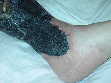
Negative-pressure wound therapy, with foam dressing, plastic film, and suction tube. (Source: Shortcut27 [CC BY-SA 3.0 (http://creativecommons.org/licenses/by-sa/3.0/)].)
BIOPHYSICAL AGENTS
The biophysical agents recommended by NPIAP are:
- Electrical stimulation
- Electromagnetic agents
- Pulsed radio frequency energy
- Ultraviolet light therapy
- Pulsatile lavage for wound cleaning and debridement
Electrical Stimulation (E-Stim)
Studies have shown that E-stim can result in significant wound healing. It is particularly useful as a treatment for wounds that have stalled in the healing process. Electrical stimulation has demonstrated several positive findings, including increased blood circulation to the wound, increase in the number on neutrophils in the wound drainage, and an increase in fibroblasts and collagen synthesis (Bryant & Nix, 2016).
Electrical stimulation therapy is applied by physical therapists via specialized electrodes that employ an electrical current to transmit energy to the tissues. The length of treatment is normally 45 to 60 minutes, with five to seven treatments each week. The following precautions are important when considering electrical stimulation as a treatment option:
- It should not be used in wounds where there is a possibility of basal or squamous cell carcinoma.
- It should not be used in the presence of osteomyelitis.
- It should not be used in patients with electrical implants (e.g., pacemakers).
- If the wound is being treated with iodine or sliver products, these must be completely removed from the wound bed before applying electrical stimulation therapy.
(Bryant & Nix, 2016; Ayello & Baranoski, 2016)
Electromagnetic Agents
This therapy uses an electromagnet to produce an electrical current to enhance wound healing. Electromagnetic fields are applied to the wound area. This treatment is also referred to as pulsed electromagnetic induction.
A Cochrane review in 2015 found there was no “strong evidence” to indicate that electromagnetic therapy was beneficial in healing pressure injuries. However, the authors pointed out that since there were only two participants in the study, further studies are needed to determine if electromagnetic therapy helps or hinders wound healing.
Pulsed Radio Frequency Energy
A pattern of low-flow electrical current is used to stimulate wound healing. Electrodes are positioned on the tissue and provide a sequence of pulses. There is a short gap between each pulse when there is no current flowing. Treatments are completed for one hour on 5 to 7 days each week, and are continued for as long as the wound is improving.
The difficulties with using any of the above forms of therapy in wound care include a lack of agreement in the wound healing community regarding the strength of evidence concerning the usefulness of the therapies in wound healing, reimbursement issues, and the lack of trained clinicians who are able to perform the therapies (Bryant & Nix, 2016).
Ultraviolet Light Therapy
A study conducted in 2008 advised against the use of ultraviolet light in the treatment of pressure injuries. However, the consortium that formulated the 2014 International Guideline for Pressure Injury Treatment, with included the NPIAP, recommended it in the short-term treatment of pressure injury (Bryant & Nix, 2016). More recent research indicates ultraviolet light treatment has the ability to reduce wound bio-burden and promote wound healing. It is effective against MRSA (Ayello & Baranoski, 2016). Ultraviolet light therapy is usually performed by physical therapists.
A frequently used procedure for administrating this therapy includes:
- Applying petrolatum to the periwound tissue
- Covering the surrounding areas with a towel or a sheet to protect against UV absorption
- Placing therapy equipment perpendicular to the skin and about one inch from the wound (newer UV therapy equipment usually has distance guards, which promotes accurate distance and accuracy of consecutive treatments)
Therapy is applied directly over the wound, with no intervening materials or substances between the wound bed and the therapy equipment.
In the past, therapy sessions lasted for 90 to 120 seconds, but current research postulates a shorter therapy time maybe more beneficial. During therapy the patient and clinician both wear UV-blocking eye protection (Bryant & Nix, 2016; Ayello & Baranoski, 2016).
RECOMBINANT PLATELET-DERIVED GROWTH FACTOR
Recombinant platelet-derived growth factors (PDGF) (becaplermin gel, brand name Regranex) play a role in regulating cell growth and division. This treatment can be considered for stages 3 and 4 pressure injuries that have delayed healing. PDGF has not been approved by the FDA for use in pressure injuries (Nix, 2016); however, it has previously been recommended for consideration in the treatment of pressure injuries by NPIAP (NPUAP/EPUAP/PPPIA, 2014) and WOCN (2016a).
PLASMA USE IN WOUND HEALING
Research in the use of platelet-rich plasma in the treatment of pressure injury is ongoing. A recent study has demonstrated a significant increase in the healing rates of stage 3 and stage 4 pressure injury healing when platelet-rich plasma was used. During this study, autologous-concentrated plasma was applied directly to the wound bed and covered with a transparent, non-adherent dressing. Further studies are needed to validate the usefulness of platelet-rich plasma in pressure injury care, to standardize treatment, and to determine the best method of application (Volalakis, 2019).
TREATMENTS NOT RECOMMENDED BY NPIAP
The following adjunct interventions are not recommended by NPIAP due to insufficient evidence of effectiveness in pressure injury treatment:
- Any other growth factors other than recombinant platelet-derived growth factor
- Bioengineered skin substitutes
- Infrared therapy
- Laser therapy
- Ultrasound
- Whirlpool
- Vibration therapy
- Topical oxygen therapy
- Hyperbaric oxygen therapy
(A discussion of these modalities is beyond the scope of this course.)
Surgical Intervention
Stages 3 and 4 pressure injuries are often difficult to heal using conventional wound healing techniques. When a pressure injury does not respond to traditional management—including debridement, infection management, and advanced wound dressings—then surgical management may be considered (NPIAP, 2016a; EPUAP/NPIAP/PPPIA, 2019). However, surgical reconstructive options may be limited due to a shortage of available tissue to use for a flap and/or impaired blood flow to the area.
Prior to surgery the patient should be in an optimal state both mentally and physically, and factors that impair healing should be minimized. The patient’s ability to tolerate the surgery and participate in the postoperative rehabilitation must be assessed prior to any surgery. Some patients may not be surgical candidates due to malnutrition, immobility, poor compliance with treatment, or chronic diseases.
Operative procedures may include skin grafts or flaps (surgical reconstruction). Myocutaneous flaps (which include both skin and muscle) are the treatment of choice for full-thickness pressure injury because they provide good protection and blood supply to the area. Immediately after surgery, the operated region must be totally and completely offloaded using a support surface that provides a high level of pressure redistribution, shear reduction, and microclimate control, with ongoing repositioning. Many hospitals use an air-fluidized support surface postoperatively. Once the surgical incision has healed, the patient will be allowed to gradually apply pressure to the area.
Surgery is a last resort due to high rates of surgical complications and pressure injury recurrence. Dehiscence of the suture line is the most common complication after surgery, ranging from 11% to 38% (WOCN, 2016). Osteomyelitis affects up to 32% of patients with pressure injuries and is the major cause of breakdown after surgery (WOCN, 2016a; EPUAP/NPIAP/PPPIA, 2019).
FLAP RECONSTRUCTION SURGERY STUDY RESULTS
A large retrospective study of flap reconstruction surgery for sacral, ischial, and trochanter pressure injuries in spinal cord injury patients at Vanderbilt University Medical Center looked at patient-specific and possible modifiable risk factors related to complications that can develop post surgery. The major complications after flap surgery included wound dehiscence and repetition of the pressure injury as great as 80%.
Findings from the study demonstrated the following:
- Age: The average age of patients who had a recurrence of pressure injury was considerably younger than those of patients who did not have a repetition of pressure injury.
- Race: African American patients had a higher incidence of pressure injury reoccurrence when compared to other racial groups.
- Location of pressure injury: The presence of an ischial pressure injury was an independent risk factor for wound dehiscence and pressure injury repetition.
- Flap choice: Flaps under higher tension had a greater risk for wound dehiscence.
- Smoking: Smoking was identified as an independent risk factor for pressure injury repetition.
- Diabetes: Diabetes was an independent risk factor for flap infection but not an independent risk factor for pressure injury repetition.
- Perioperative blood transfusions: Perioperative blood transfusions were linked to postoperative complications, including flap infection, wound dehiscence, and pressure injury repletion.
- Wound complexity: Larger wounds and prolonged operative periods resulted in greater incidence of flap infection, and extended operative times were related to a greater incidence of wound dehiscence.
(Bamba et al., 2017)