PATHOPHYSIOLOGY OF THE LUNG
Normal Lungs
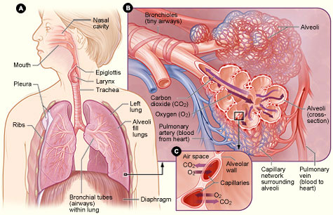
Figure A: Locations of the respiratory structures in the body.
Figure B: Enlarged image of airways, alveoli, and their capillaries in the lungs.
Figure C: Location of gas exchange between the capillaries and alveoli.
(Source: National Institutes of Health.)
LUNG STRUCTURE
The lungs are a pair of cone-shaped organs in the thoracic cavity. Each lung has three surfaces: anterior, posterior, and inferior. The right lung is larger and weighs more than the left lung, and it has three lobes. The left lung has two lobes and a space for the heart, called a cardiac notch (Physiopedia, 2020).
Each type of lung tissue is differentiated from the others. Mature, differentiated cells are distinguished from other types by a unique appearance as well as function.
Pleura
Both lungs are surrounded by a serous pleural sac comprised of two continuous membranes. There are four components of the parietal pleura: 1) the costal pleura line the thoracic wall; 2) the mediastinal pleura line the lateral mediastinum; 3) the diaphragmatic pleura line the superior diaphragm and both sides of the mediastinum; and 4) the cervical pleura extends to the neck and covers the apices of the lungs (Physiopedia, 2020).
Pleural Cavity
The pleural cavity is a potential space between the parietal and visceral layers of the pleura. Serous fluid in the pleural cavity lubricates the two surfaces of the pleura so that they can move easily over each other during respirations. Surface tension in the pleural cavity causes the lungs to stick to the thoracic wall and leads to a close proximity of the lung surfaces with the chest all, which supports more inflation of the alveoli during inspiration (Physiopedia, 2020).
Lung Fissures
Each lung is divided by fissures (connective tissue walls) into separate lobes. Both lungs have oblique fissures dividing the lobes, and the right lung has an additional, transverse fissure that forms the third lobe (Physiopedia, 2020).
Bronchial Tree
At the top of the bronchial tree is the trachea. The trachea divides into the right and left primary bronchus or main bronchi. Each bronchus enters the lung at a notch called the hilum. The right main bronchus is less angled and has a larger diameter than the left and exits the trachea at a higher level than the left main bronchus. Anything foreign that enters the bronchial tree will tend to enter into the right main bronchus rather than the left, primarily due to size and position. The main bronchi further divide into secondary and then tertiary bronchi. The tertiary bronchi supply the bronchopulmonary segments with air (Physiopedia, 2020).
Bronchopulmonary Segments
The bronchopulmonary segments are the largest divisions of a lobe. The tertiary bronchi further subdivide into smaller bronchioles. Each of these continue to subdivide into 50–80 even smaller bronchioles. Eventually, the terminal bronchioles form the approximately 300 million alveolar ducts and sacs, where the exchange of oxygen and carbon dioxide takes place during respirations (Physiopedia, 2020).
Bronchopulmonary Segment Histology
The trachea is surrounded by cartilage rings, smooth muscles, and elastic fibers. Goblet cells for mucus production are lined with ciliated epithelia shaped like columns, which aid in secretion removal. The smaller bronchioles have elastic tissues and smooth muscle fibers. The alveoli are fashioned of thin squamous epithelial cells. The gas exchange of oxygen and carbon dioxide takes place between these cells (Physiopedia, 2020).
LUNG FUNCTION
Respirations allow the exchange of oxygen (O2) and carbon dioxide (CO2). Oxygen is inhaled from the atmosphere to the alveoli, where the gas exchange takes place. In the alveolar capillary membranes, venous blood becomes oxygenated arterial blood that returns to the body. The process of oxygenation takes place via ventilation, perfusion, and diffusion.
Ventilation is the movement of gases in and out of the lungs. Perfusion is the cardiovascular system pumping oxygenated blood to the tissues and deoxygenated blood back to the lungs, where it will be reoxygenated. Diffusion is the exchange of respiratory gases in the alveolar capillary membranes. Surfactant is a chemical that serves to keep the alveolar sacs open so that they may fill with inhaled air.
The accessory muscles of ventilation (the scalene, sternocleidomastoid, pectoralis major, trapezius, and external intercostal muscles) can increase the lungs’ volumes. Excessive use of these muscles causes fatigue. Compliance is the ability of the lungs to expand or distend as a result of intra-alveolar pressure. Airway resistance is the increase in pressure as the diameter of the airway becomes progressively smaller along the passage of air from the mouth or nose to the alveoli. Tumors in the airway will also increase airway resistance. The increased use of accessory muscles, decreased lung compliance, and increased airway resistance result in the increased work of breathing, causing increased energy expenditure and oxygen consumption (Potter et al., 2019).
Lungs with Lung Cancer
The majority of primary lung tumors originate in mutated epithelial tissue that was previously normal tissue. Mutated tissue differs in behavior and appearance from normal tissue. In a normal cell, it is the DNA that controls the growth and behavior of new cells. Cancer can cause the DNA to create cells that are abnormal in appearance and to reproduce more rapidly (NCCN, 2019).
There are several different types of lung cancers, categorized based on the cellular and molecular characteristics of the cancer cells present (histopathological class). It is vitally important to know the histopathology of the lung cancer to correctly diagnose and treat the cancer (Siddiqui et al., 2021). (See also “Types and Staging of Lung Cancer” later in this course.)
Carcinogenic factors cause the mutations, depending on genetic factors. The slow tumor development is fostered by epidermal growth factors. It takes 8–10 years for a lung tumor to grow large enough to be detected by X-ray. Lung cancer tumors usually arise in the segmental bronchi or the upper lobes (Harding et al., 2020).